Copyright
©The Author(s) 2023.
World J Clin Cases. Aug 26, 2023; 11(24): 5797-5803
Published online Aug 26, 2023. doi: 10.12998/wjcc.v11.i24.5797
Published online Aug 26, 2023. doi: 10.12998/wjcc.v11.i24.5797
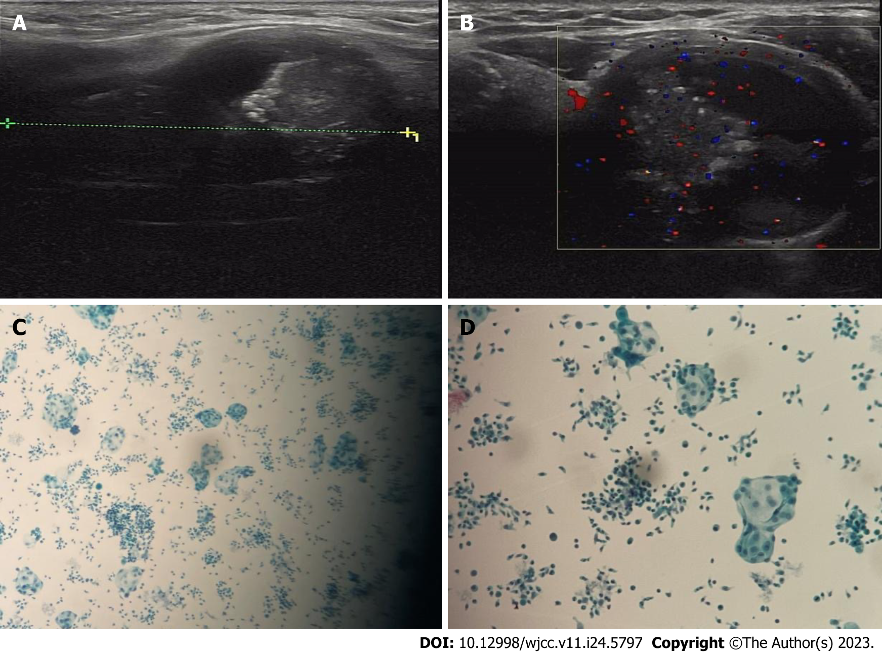
Figure 1 Neck ultrasonography.
A and B: Ultrasonography image of the neck showed a mixed echoic and horizontal nodule measuring approximately 5.1 cm × 3.1 cm × 2.9 cm in the left thyroid gland; C and D: Fine needle aspiration revealed a mass of hyperplastic glandular epithelial cells and infiltration of short spindle cells.
- Citation: Hu J, Wang F, Xue W, Jiang Y. Papillary thyroid carcinoma with nodular fasciitis-like stroma - an unusual variant with distinctive histopathology: A case report. World J Clin Cases 2023; 11(24): 5797-5803
- URL: https://www.wjgnet.com/2307-8960/full/v11/i24/5797.htm
- DOI: https://dx.doi.org/10.12998/wjcc.v11.i24.5797


