Copyright
©The Author(s) 2023.
World J Clin Cases. Aug 26, 2023; 11(24): 5772-5779
Published online Aug 26, 2023. doi: 10.12998/wjcc.v11.i24.5772
Published online Aug 26, 2023. doi: 10.12998/wjcc.v11.i24.5772
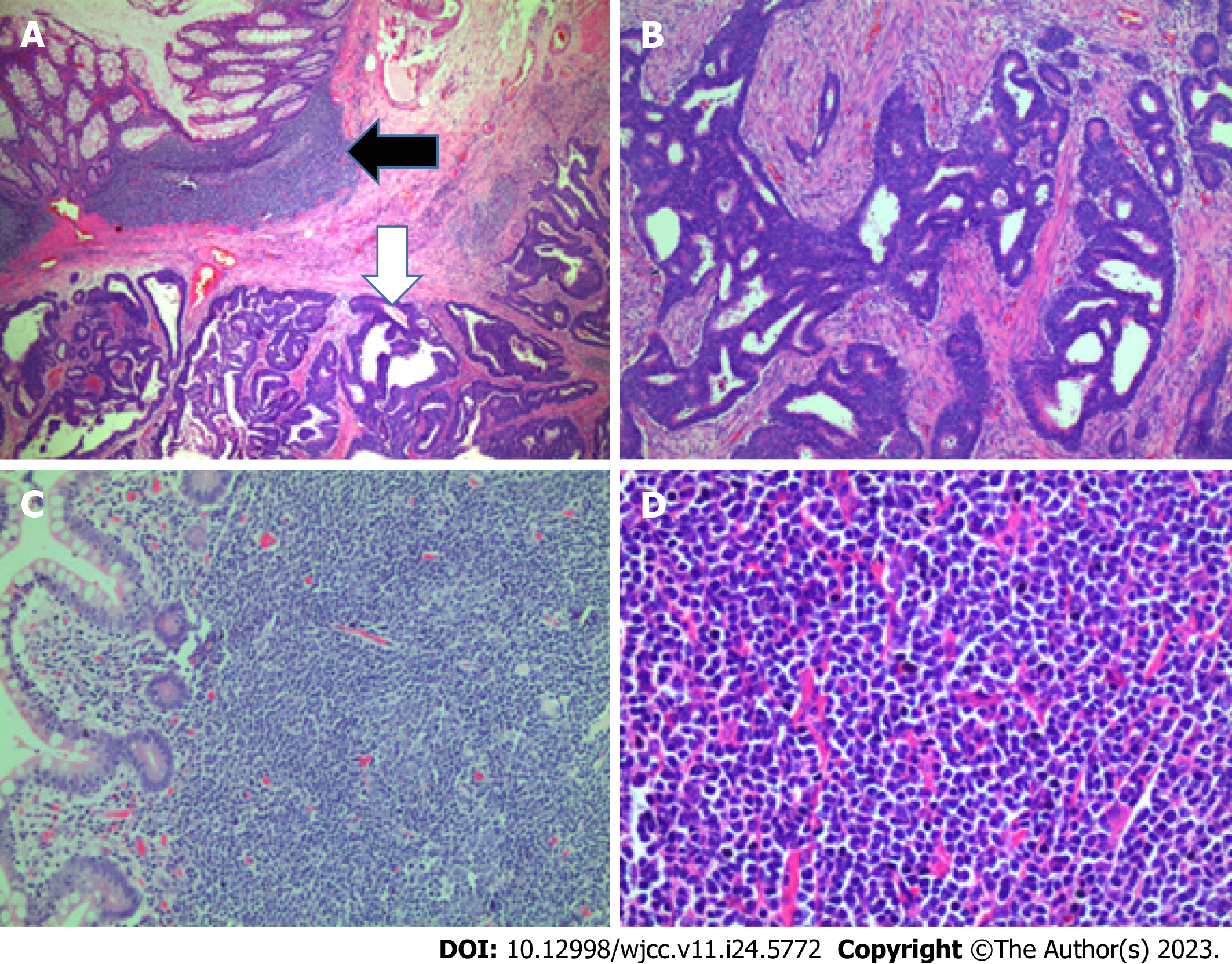
Figure 3 The microscopic appearance of the lesion.
A: A loupe view of a rectal cross section shows both adenocarcinoma tumors (white arrow) and lymphoma tumors (black arrow) [hematoxylin and eosin (H&E) staining, 4×]; B: An infiltrating moderately differentiated adenocarcinoma of the rectum (H&E staining, 10×); C: Mucosal and submucosal nodules of lymphoma on polyps (H&E staining, 20×); D: H&E staining of lymphoma (magnification 40×).
- Citation: Vu KV, Trong NV, Khuyen NT, Huyen Nga D, Anh H, Tien Trung N, Trung Thong P, Minh Duc N. Synchronous rectal adenocarcinoma and intestinal mantle cell lymphoma: A case report. World J Clin Cases 2023; 11(24): 5772-5779
- URL: https://www.wjgnet.com/2307-8960/full/v11/i24/5772.htm
- DOI: https://dx.doi.org/10.12998/wjcc.v11.i24.5772


