Copyright
©The Author(s) 2023.
World J Clin Cases. Aug 26, 2023; 11(24): 5762-5771
Published online Aug 26, 2023. doi: 10.12998/wjcc.v11.i24.5762
Published online Aug 26, 2023. doi: 10.12998/wjcc.v11.i24.5762
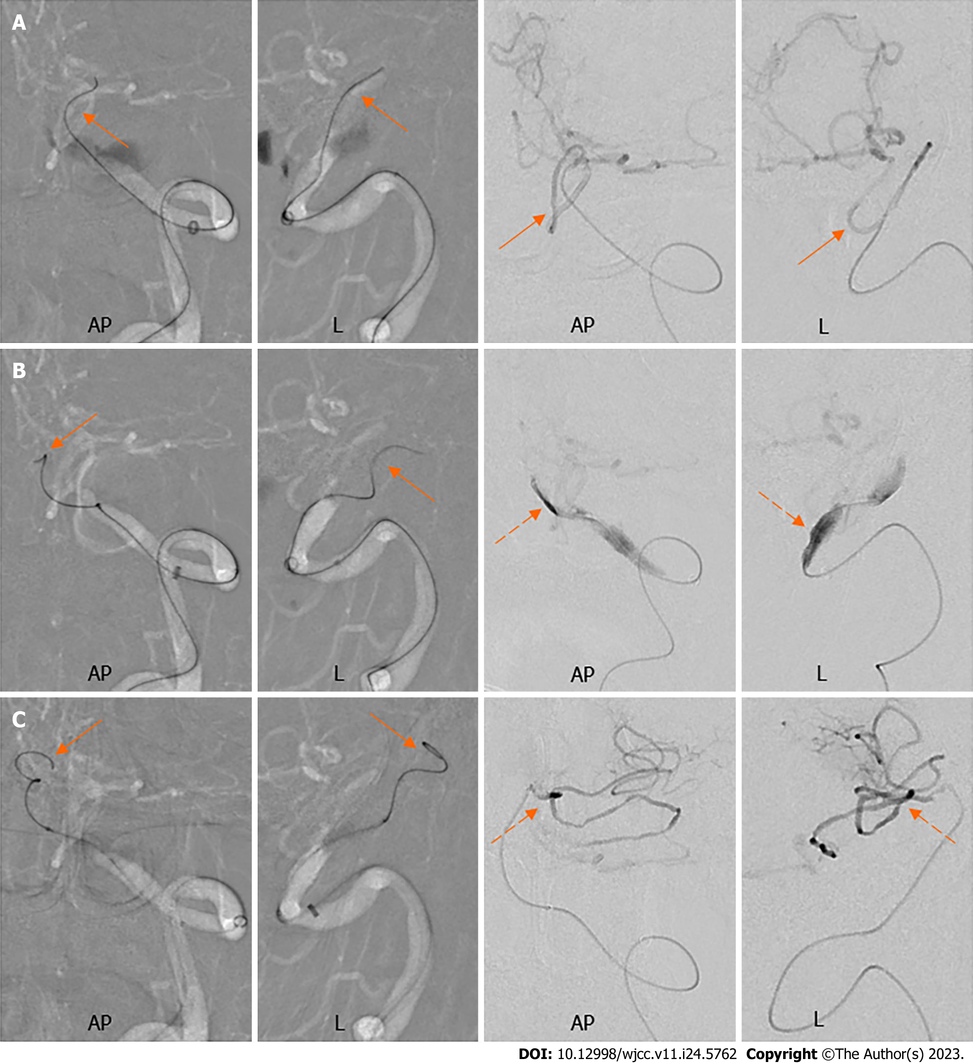
Figure 4 The course of microwire in the subintima and microcatheter angiography.
A: The microwire entered the posterior inferior cerebellar artery (orange arrows); B: The microwire advanced in a sigmoid curve in the subintima (orange arrows) and the microcatheter angiography identified the subintimal dissection (orange dashed arrows); C: The subintimal microwire kept advancing in a spiral shape (orange arrows) and the microcatheter angiography showed the microwire entered the anterior inferior cerebellar artery (orange dashed arrows). AP: Anterior-posterior; L: Lateral.
- Citation: Fu JF, Zhang XL, Lee SY, Zhang FM, You JS. Subintimal recanalization for non-acute occlusion of intracranial vertebral artery in an emergency endovascular procedure: A case report. World J Clin Cases 2023; 11(24): 5762-5771
- URL: https://www.wjgnet.com/2307-8960/full/v11/i24/5762.htm
- DOI: https://dx.doi.org/10.12998/wjcc.v11.i24.5762


