Copyright
©The Author(s) 2023.
World J Clin Cases. Aug 26, 2023; 11(24): 5762-5771
Published online Aug 26, 2023. doi: 10.12998/wjcc.v11.i24.5762
Published online Aug 26, 2023. doi: 10.12998/wjcc.v11.i24.5762
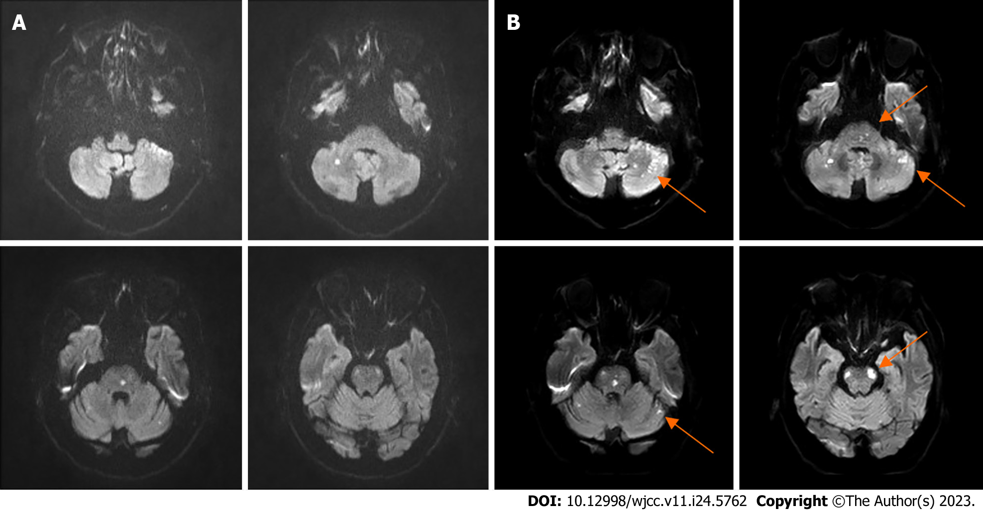
Figure 1 The brain diffusion-weighted magnetic resonance imaging at different time intervals.
A: Diffusion-weighted magnetic resonance imaging (DW-MRI) at initial admission; B: DW-MRI on day 4 of admission showed multiple infarcts enlarged than before (orange arrows).
- Citation: Fu JF, Zhang XL, Lee SY, Zhang FM, You JS. Subintimal recanalization for non-acute occlusion of intracranial vertebral artery in an emergency endovascular procedure: A case report. World J Clin Cases 2023; 11(24): 5762-5771
- URL: https://www.wjgnet.com/2307-8960/full/v11/i24/5762.htm
- DOI: https://dx.doi.org/10.12998/wjcc.v11.i24.5762


