Copyright
©The Author(s) 2023.
World J Clin Cases. Aug 26, 2023; 11(24): 5692-5699
Published online Aug 26, 2023. doi: 10.12998/wjcc.v11.i24.5692
Published online Aug 26, 2023. doi: 10.12998/wjcc.v11.i24.5692
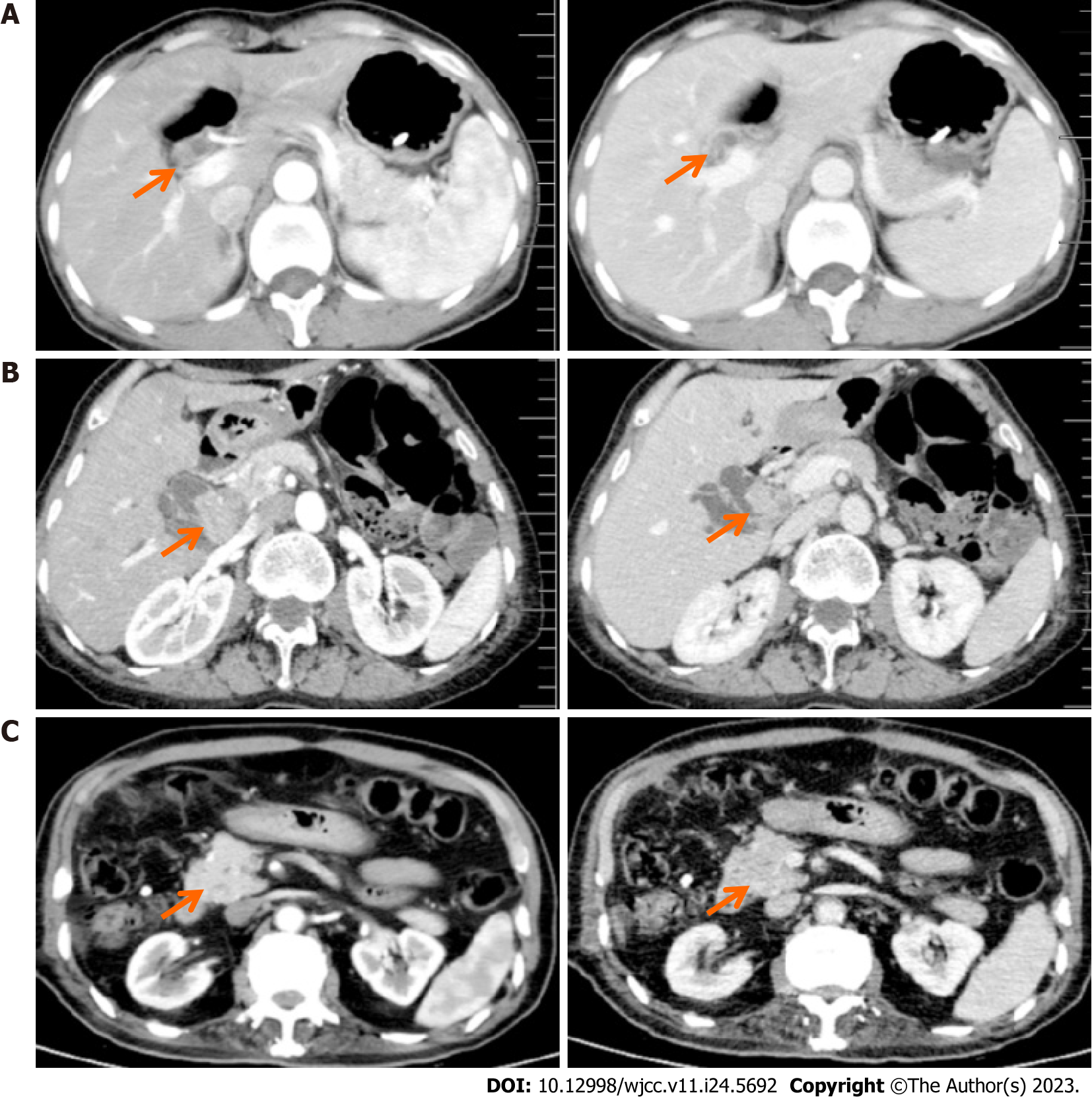
Figure 3 Contrast-enhanced computed tomography scan of extrahepatic biliary adenoma.
A-C: Contrast-enhanced computed tomography scan shows dilatation of the intrahepatic or extra-hepatic bile duct with a slightly enhancement of the mass in left hepatic duct (A), hilar bile duct (B) and distal common bile duct (C). The arrow symbols point to adenomas.
- Citation: Li W, Tao J, Song XG, Hou MR, Qu K, Gu JT, Yan XP, Yao BW, Qin YF, Dong FF, Sha HC. Clinical study of extrahepatic biliary adenoma. World J Clin Cases 2023; 11(24): 5692-5699
- URL: https://www.wjgnet.com/2307-8960/full/v11/i24/5692.htm
- DOI: https://dx.doi.org/10.12998/wjcc.v11.i24.5692


