Copyright
©The Author(s) 2023.
World J Clin Cases. Aug 16, 2023; 11(23): 5622-5627
Published online Aug 16, 2023. doi: 10.12998/wjcc.v11.i23.5622
Published online Aug 16, 2023. doi: 10.12998/wjcc.v11.i23.5622
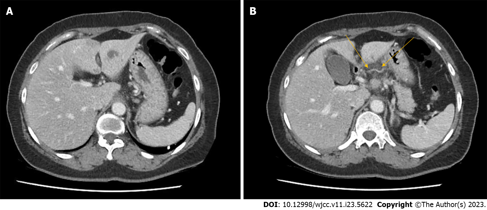
Figure 2 Abdominal computed tomography scan results of the patient.
A: Thin linear radiopaque lesion with adjacent low attenuating lesion in the S3 segment of liver; B: Mild infiltration at lesser curvature side of stomach antrum (orange arrows), suggesting foreign body penetration from stomach to liver.
- Citation: Park Y, Han HS, Yoon YS, Cho JY, Lee B, Kang M, Kim J, Lee HW. Pyogenic liver abscess secondary to gastric perforation of an ingested toothpick: A case report. World J Clin Cases 2023; 11(23): 5622-5627
- URL: https://www.wjgnet.com/2307-8960/full/v11/i23/5622.htm
- DOI: https://dx.doi.org/10.12998/wjcc.v11.i23.5622


