Copyright
©The Author(s) 2023.
World J Clin Cases. Aug 16, 2023; 11(23): 5602-5609
Published online Aug 16, 2023. doi: 10.12998/wjcc.v11.i23.5602
Published online Aug 16, 2023. doi: 10.12998/wjcc.v11.i23.5602
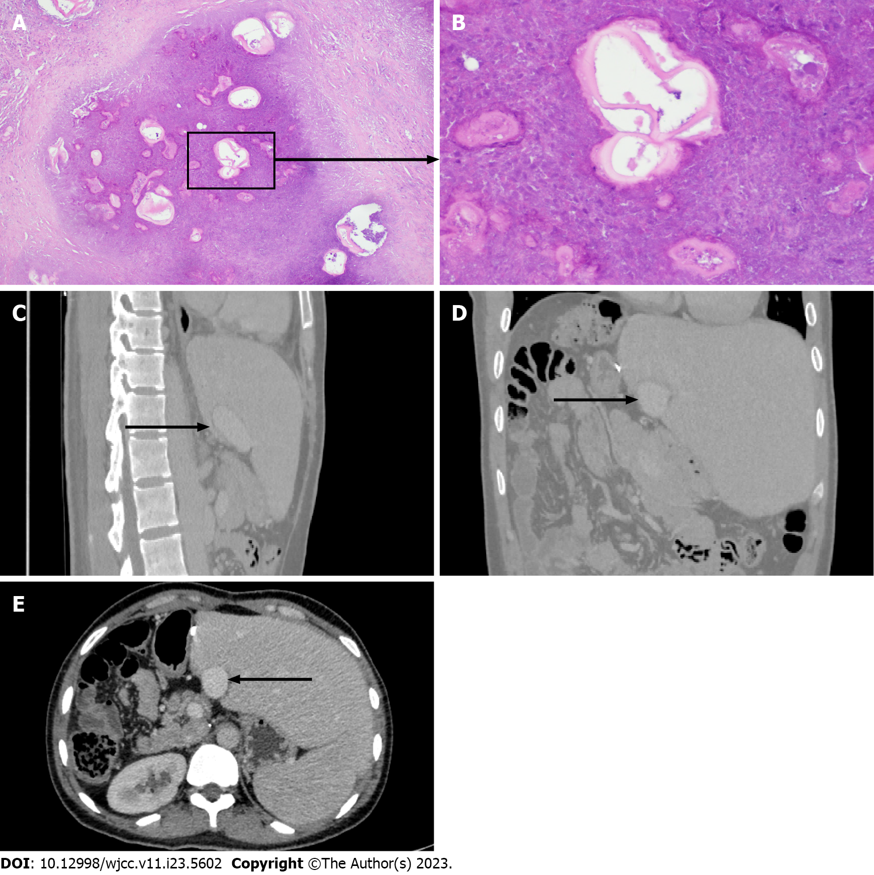
Figure 2 Pathology of alveolar echinococcosis and long-term follow up 7 years after surgery.
A: Lesions of hepatic alveolar echinococcosis (AE) (hematoxylin and eosin, ×10); B: Lesions of hepatic AE (hematoxylin and eosin, ×40). Long-term follow-up abdominal computed tomography (CT) shows the morphology of the reconstructed inferior vena cava (IVC) in the coronal plane, sagittal plane, and cross-section at 84 mo post-surgery; C-E: The black arrow indicates the reconstructed IVC.
- Citation: Humaerhan J, Jiang TM, Aji T, Shao YM, Wen H. Complex inferior vena cava reconstruction during ex vivo liver resection and autotransplantation: A case report. World J Clin Cases 2023; 11(23): 5602-5609
- URL: https://www.wjgnet.com/2307-8960/full/v11/i23/5602.htm
- DOI: https://dx.doi.org/10.12998/wjcc.v11.i23.5602


