Copyright
©The Author(s) 2023.
World J Clin Cases. Aug 16, 2023; 11(23): 5573-5579
Published online Aug 16, 2023. doi: 10.12998/wjcc.v11.i23.5573
Published online Aug 16, 2023. doi: 10.12998/wjcc.v11.i23.5573
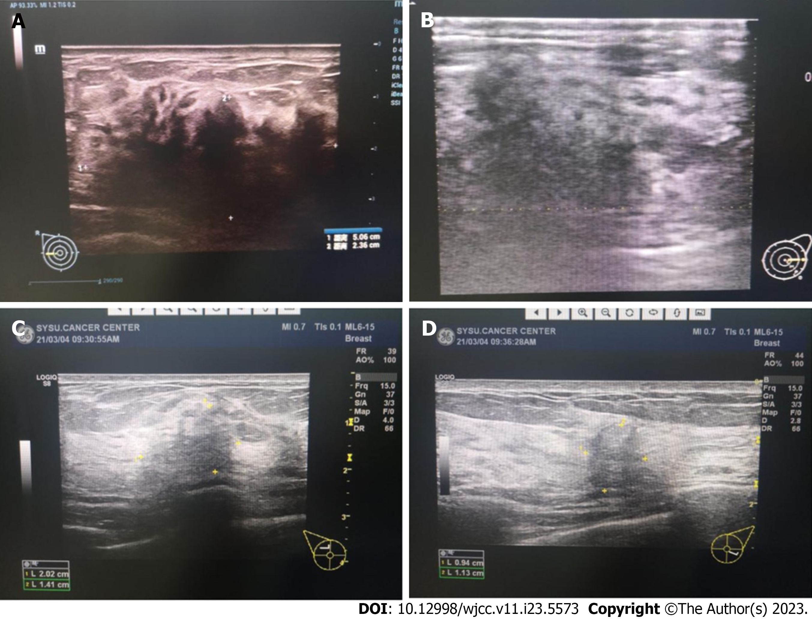
Figure 3 Breast color Doppler ultrasound images.
A and B: Images of the breasts at admission showing irregular masses on both sides, with incomplete margins, angulation, and burrs; C and D: Images of the breasts after six cycles of chemotherapy showing that the masses had significantly decreased in size, with uneven internal echo and no point-like strong echo.
- Citation: Lei YY, Li DM. Nasopharyngeal carcinoma with synchronous breast metastasis: A case report. World J Clin Cases 2023; 11(23): 5573-5579
- URL: https://www.wjgnet.com/2307-8960/full/v11/i23/5573.htm
- DOI: https://dx.doi.org/10.12998/wjcc.v11.i23.5573


