Copyright
©The Author(s) 2023.
World J Clin Cases. Aug 16, 2023; 11(23): 5479-5493
Published online Aug 16, 2023. doi: 10.12998/wjcc.v11.i23.5479
Published online Aug 16, 2023. doi: 10.12998/wjcc.v11.i23.5479
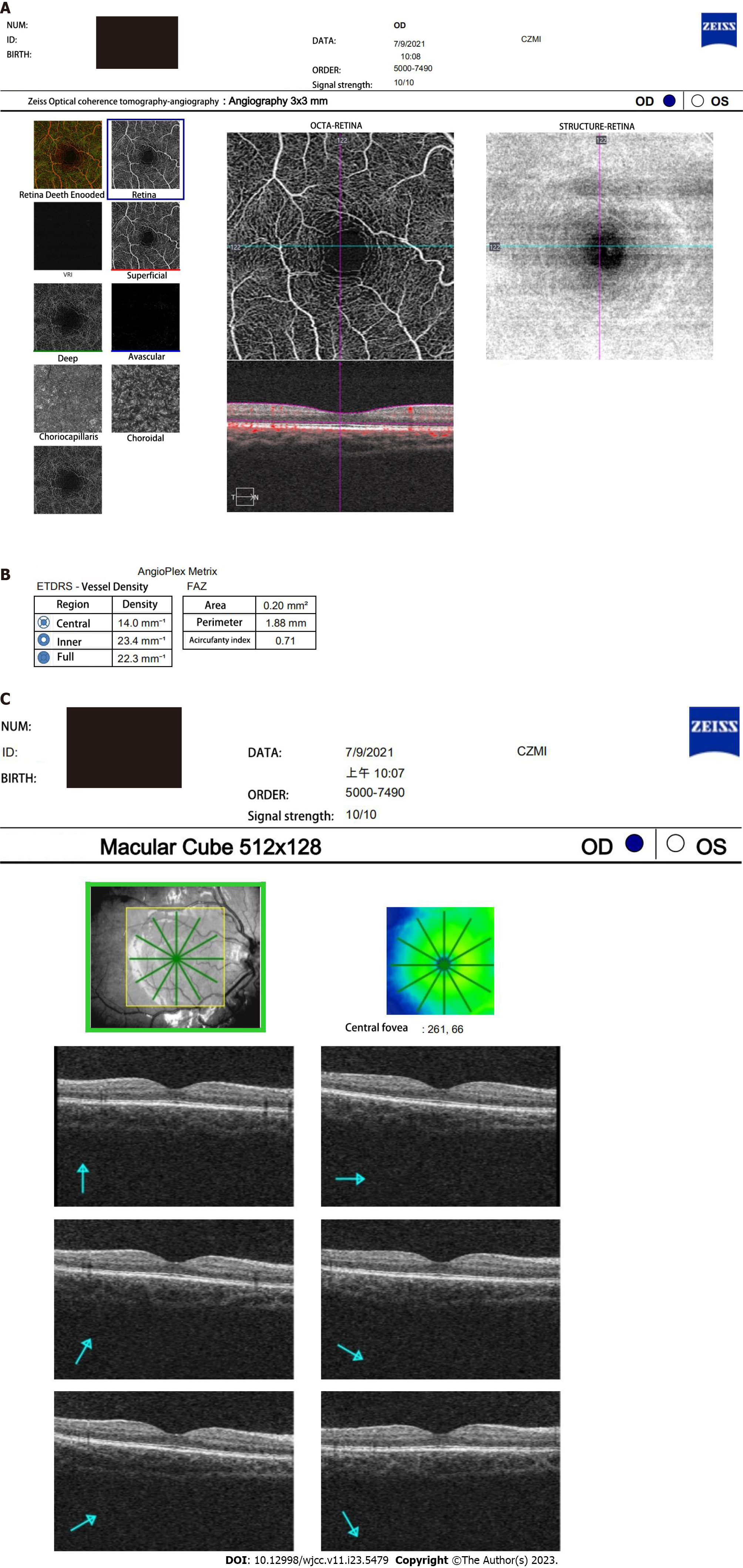
Figure 2 The optical coherence tomography angiography images obtained by participant.
A: Optical coherence tomography angiography (OCTA) images of superficial capillary plexus (SCP) in the macula; B: OCTA image showing 3 mm × 3 mm scanning model in the SCP of the macula. OCTA image showing foveal avascular zone area obtained automatically by OCTA software; C: OCTA image showing 512 × 128 scanning model in the macular cube. FAZ: Foveal avascular zone.
- Citation: Sun KX, Xiang YG, Zhang T, Yi SL, Xia JY, Yang X, Zheng SJ, Ji Y, Wan WJ, Hu K. Evaluation of childhood developing via optical coherence tomography-angiography in Qamdo, Tibet, China: A prospective cross-sectional, school-based study. World J Clin Cases 2023; 11(23): 5479-5493
- URL: https://www.wjgnet.com/2307-8960/full/v11/i23/5479.htm
- DOI: https://dx.doi.org/10.12998/wjcc.v11.i23.5479


