Copyright
©The Author(s) 2023.
World J Clin Cases. Aug 16, 2023; 11(23): 5430-5439
Published online Aug 16, 2023. doi: 10.12998/wjcc.v11.i23.5430
Published online Aug 16, 2023. doi: 10.12998/wjcc.v11.i23.5430
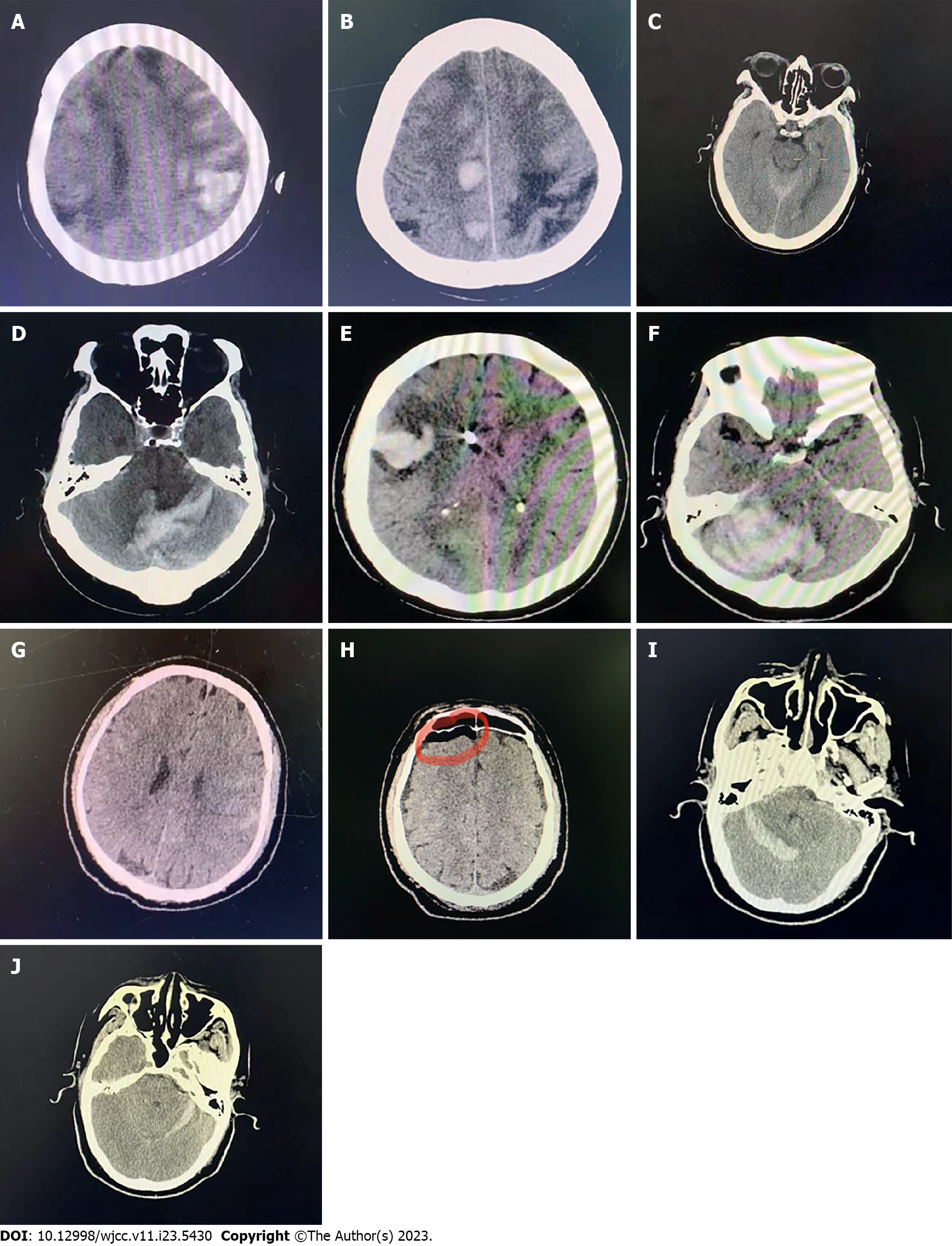
Figure 1 Imaging manifestations of intracranial hemorrhage after spinal surgery.
A: Case 1, bilateral parietal and frontal lobe hemorrhage and subarachnoid hemorrhage for the first time; B: Case 1, left parietal and frontal lobar hemorrhage for the second time; C: Case 2, subarachnoid hemorrhage and ventricular hemorrhage; D: Case 3, cerebellar hemorrhage; E-F: Case 4, right frontal lobe hemorrhage and right cerebellar hemorrhage; G: Case 5, subarachnoid hemorrhage; H: Case 5, intracranial pneumatosis; I-J: Case 6, bilateral cerebellar hemorrhage.
- Citation: Yan X, Yan LR, Ma ZG, Jiang M, Gao Y, Pang Y, Wang WW, Qin ZH, Han YT, You XF, Ruan W, Wang Q. Clinical characteristics and risk factors of intracranial hemorrhage after spinal surgery. World J Clin Cases 2023; 11(23): 5430-5439
- URL: https://www.wjgnet.com/2307-8960/full/v11/i23/5430.htm
- DOI: https://dx.doi.org/10.12998/wjcc.v11.i23.5430


