Copyright
©The Author(s) 2023.
World J Clin Cases. Jul 26, 2023; 11(21): 5122-5128
Published online Jul 26, 2023. doi: 10.12998/wjcc.v11.i21.5122
Published online Jul 26, 2023. doi: 10.12998/wjcc.v11.i21.5122
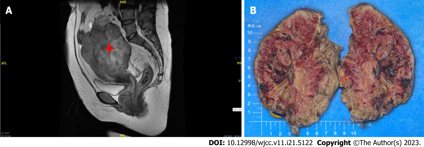
Figure 2 Magnetic resonance imaging findings and macroscopic observation for case 2.
A: Magnetic resonance imaging documented a solid mass 10.3 cm × 9.2 cm in dimensions, uniform density, regular margin, and unclear boundary in the right adnexal area ureter; B: The right ovary was markedly swollen, and it was totally displaced by a solid mass. The cut surface was red; spongy areas occupied a substantial portion.
- Citation: Zhou Y, Sun YW, Liu XY, Shen DH. Primary ovarian angiosarcoma: Two case reports and review of literature. World J Clin Cases 2023; 11(21): 5122-5128
- URL: https://www.wjgnet.com/2307-8960/full/v11/i21/5122.htm
- DOI: https://dx.doi.org/10.12998/wjcc.v11.i21.5122


