Copyright
©The Author(s) 2023.
World J Clin Cases. Jul 26, 2023; 11(21): 5047-5055
Published online Jul 26, 2023. doi: 10.12998/wjcc.v11.i21.5047
Published online Jul 26, 2023. doi: 10.12998/wjcc.v11.i21.5047
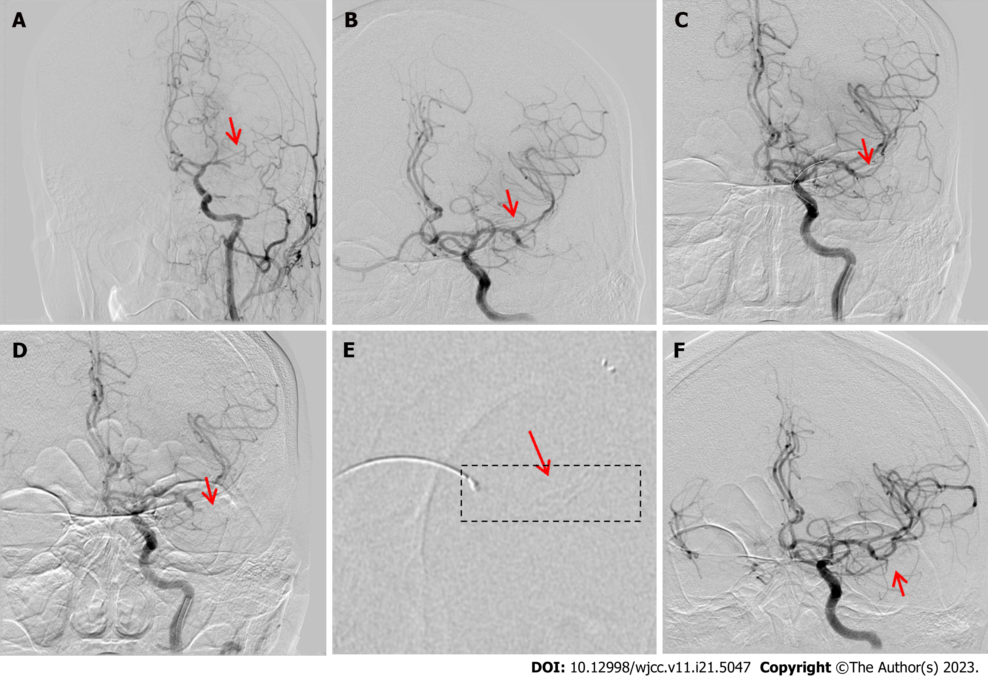
Figure 2 Operative procedure: Patient 2.
A: Digital subtraction angiography (DSA) revealing left middle cerebral artery (LMCA)-M1 segment occlusion (arrow); B: DSA revealing good recanalization of the M2 segment upper (arrow) and lower branch occlusions; C: The stent was deployed and retrieved without stent imaging (arrow); D: Angiogram revealing no recanalization postoperatively (arrow); E: The stent was redeployed at the distal M2 segment with a partial development of stent imaging; F: DSA after stent deployment revealing good recanalization of the LMCA-M2 segment (arrow).
- Citation: Yao QY, Fu ML, Zhao Q, Zheng XM, Tang K, Cao LM. Image-based visualization of stents in mechanical thrombectomy for acute ischemic stroke: Preliminary findings from a series of cases. World J Clin Cases 2023; 11(21): 5047-5055
- URL: https://www.wjgnet.com/2307-8960/full/v11/i21/5047.htm
- DOI: https://dx.doi.org/10.12998/wjcc.v11.i21.5047


