Copyright
©The Author(s) 2023.
World J Clin Cases. Jul 16, 2023; 11(20): 4944-4955
Published online Jul 16, 2023. doi: 10.12998/wjcc.v11.i20.4944
Published online Jul 16, 2023. doi: 10.12998/wjcc.v11.i20.4944
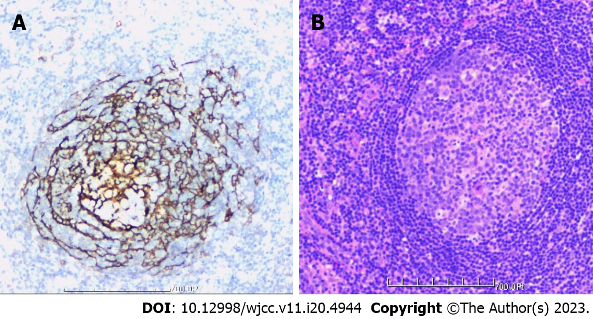
Figure 10 Postoperative pathological examination of tissue specimens.
The tissue was primarily hyperplastic and degenerative fibrous tissue and degenerative bone tissue, with flaky distribution, partial necrosis of bone tissue, interstitial vascular hyperplasia and dilatation with inflammatory cell infiltration, patchy areas of necrosis (pink), and a significant amount of multinucleated giant-cell infiltration. Plasma cell CD38 (focus +), CD138, lymphocyte LCA (+), Ki-67 approximately 10% graded CK (-), EMA (-), Smur100 (+), CD68 (histiocyte +), CD1a (histiocyte +), SMA (vascular +), and CD34 (vascular +) were all detected in the tissue by immunohistochemistry. The diagnosis of EG was supported by histology, immunohistochemical staining, and other factors. A: Immunohistochemistry; B: Hematoxylin and eosin staining.
- Citation: Tu CQ, Chen ZD, Yao XT, Jiang YJ, Zhang BF, Lin B. Posterior pedicle screw fixation combined with local steroid injections for treating axial eosinophilic granulomas and atlantoaxial dislocation: A case report. World J Clin Cases 2023; 11(20): 4944-4955
- URL: https://www.wjgnet.com/2307-8960/full/v11/i20/4944.htm
- DOI: https://dx.doi.org/10.12998/wjcc.v11.i20.4944


