Copyright
©The Author(s) 2023.
World J Clin Cases. Jul 16, 2023; 11(20): 4937-4943
Published online Jul 16, 2023. doi: 10.12998/wjcc.v11.i20.4937
Published online Jul 16, 2023. doi: 10.12998/wjcc.v11.i20.4937
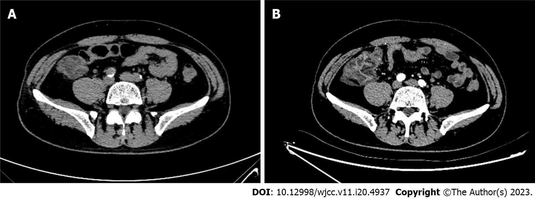
Figure 2 Abdominal computed tomography scan (axial plane).
A: At 9 h post- coronary angiography (CAG), computed tomography revealed thickened right colonic wall accompanied by multiple exudative changes indicating inflammatory lesions; B: On the 3rd day after CAG, edema and inflammatory exudate became more serious.
- Citation: Qiu H, Li WP. Contrast-induced ischemic colitis following coronary angiography: A case report. World J Clin Cases 2023; 11(20): 4937-4943
- URL: https://www.wjgnet.com/2307-8960/full/v11/i20/4937.htm
- DOI: https://dx.doi.org/10.12998/wjcc.v11.i20.4937


