Copyright
©The Author(s) 2023.
World J Clin Cases. Jul 16, 2023; 11(20): 4912-4919
Published online Jul 16, 2023. doi: 10.12998/wjcc.v11.i20.4912
Published online Jul 16, 2023. doi: 10.12998/wjcc.v11.i20.4912
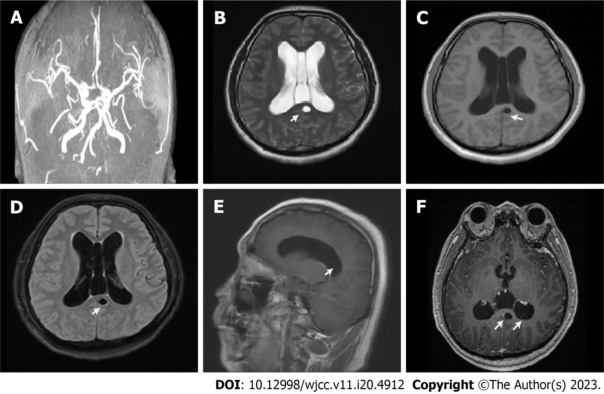
Figure 1 Imaging examinations results.
A: Magnetic resonance arteriography image revealing partial vascular stenosis and weakened blood flow signal; B-D: Cerebral magnetic resonance imaging (MRI) showing a cystic lesion, an expanded ventricular system and hydrocephalus. T1-weighted image showing a low signal of the corpus callosum (B); T2-weighted image showing a high signal behind the corpus callosum (C); Fast fluid attenuated inversion recovery sequence of brain MRI revealing a cystic lesion in the corpus callosum (D); E and F: Cerebral MRI image with contrast showing several small hyperintense lesions involving the brain parenchyma and ventricular system, with subtle ring enhancement of gadolinium.
- Citation: Xu WB, Fu JJ, Yuan XJ, Xian QJ, Zhang LJ, Song PP, You ZQ, Wang CT, Zhao QG, Pang F. Metagenomic next-generation sequencing in the diagnosis of neurocysticercosis: A case report. World J Clin Cases 2023; 11(20): 4912-4919
- URL: https://www.wjgnet.com/2307-8960/full/v11/i20/4912.htm
- DOI: https://dx.doi.org/10.12998/wjcc.v11.i20.4912


