Copyright
©The Author(s) 2023.
World J Clin Cases. Jan 16, 2023; 11(2): 417-425
Published online Jan 16, 2023. doi: 10.12998/wjcc.v11.i2.417
Published online Jan 16, 2023. doi: 10.12998/wjcc.v11.i2.417
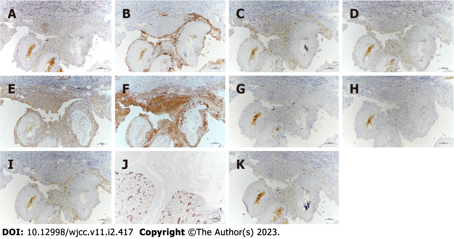
Figure 3 Immunoprofile of renal angiomyolipoma with cystic degeneration.
A-F: Tumor cells exhibited positivity for neuron-specific enolase (A), human melanoma black-45 (B), Melan-A (C), S-100 (D), smooth muscle actin (E), and calponin (F); G-J: They did not express estrogen receptor (G), progesterone receptor (H), CD68 (I), or cytokeratin (J); K: The Ki-67 labeling index was less than 5%. Original magnification × 200.
- Citation: Lu SQ, Lv W, Liu YJ, Deng H. Fat-poor renal angiomyolipoma with prominent cystic degeneration: A case report and review of the literature. World J Clin Cases 2023; 11(2): 417-425
- URL: https://www.wjgnet.com/2307-8960/full/v11/i2/417.htm
- DOI: https://dx.doi.org/10.12998/wjcc.v11.i2.417


