Copyright
©The Author(s) 2023.
World J Clin Cases. Jan 16, 2023; 11(2): 417-425
Published online Jan 16, 2023. doi: 10.12998/wjcc.v11.i2.417
Published online Jan 16, 2023. doi: 10.12998/wjcc.v11.i2.417
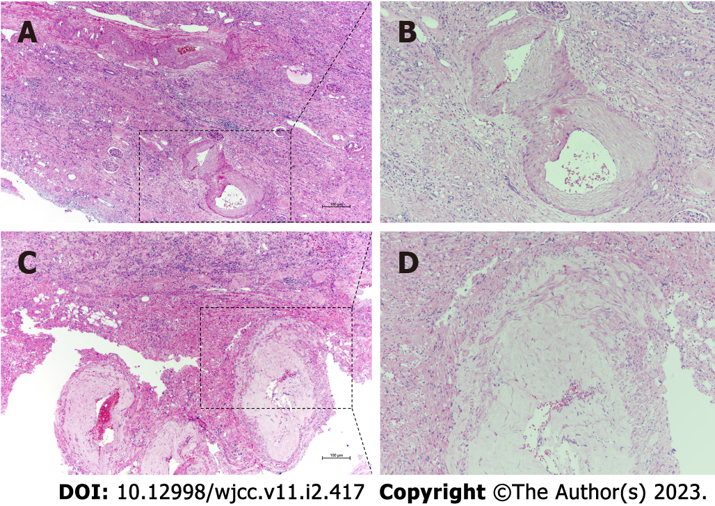
Figure 2 Histopathological findings of renal angiomyolipoma with cystic degeneration.
A: Hematoxylin and eosin staining showed tortuous and ectatic tumor vessels with uneven and thick wall in the critical renal parenchyma; B: Randomly arranged smooth muscle-like spindle cells with nuclei of different sizes. Original magnification × 200 and × 400 (insets).
- Citation: Lu SQ, Lv W, Liu YJ, Deng H. Fat-poor renal angiomyolipoma with prominent cystic degeneration: A case report and review of the literature. World J Clin Cases 2023; 11(2): 417-425
- URL: https://www.wjgnet.com/2307-8960/full/v11/i2/417.htm
- DOI: https://dx.doi.org/10.12998/wjcc.v11.i2.417


