Copyright
©The Author(s) 2023.
World J Clin Cases. Jan 16, 2023; 11(2): 417-425
Published online Jan 16, 2023. doi: 10.12998/wjcc.v11.i2.417
Published online Jan 16, 2023. doi: 10.12998/wjcc.v11.i2.417
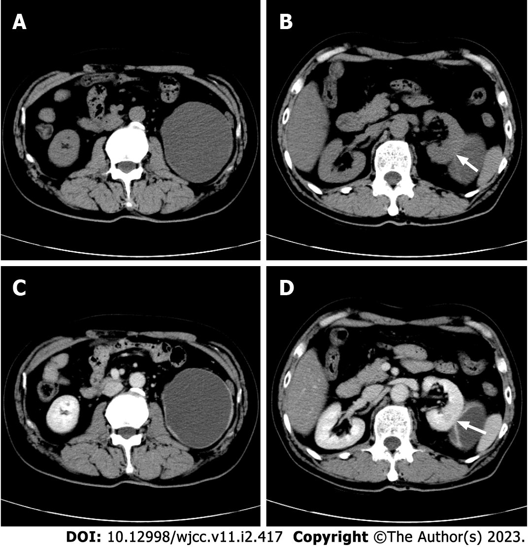
Figure 1 Computed tomography of the kidney with cystic degeneration.
A: Plain computed tomography (CT) revealed an 8.6 cm × 7.4 cm, oval to round, mixed hypodense and isodense, cystic exophytic lesion with mainly liquid density; B: A smooth linear septum was observed (arrow); C: Enhanced scan revealed enhancement of the nodules and irregular wall; D: The smooth linear septum (arrow).
- Citation: Lu SQ, Lv W, Liu YJ, Deng H. Fat-poor renal angiomyolipoma with prominent cystic degeneration: A case report and review of the literature. World J Clin Cases 2023; 11(2): 417-425
- URL: https://www.wjgnet.com/2307-8960/full/v11/i2/417.htm
- DOI: https://dx.doi.org/10.12998/wjcc.v11.i2.417


