Copyright
©The Author(s) 2023.
World J Clin Cases. Jan 16, 2023; 11(2): 255-267
Published online Jan 16, 2023. doi: 10.12998/wjcc.v11.i2.255
Published online Jan 16, 2023. doi: 10.12998/wjcc.v11.i2.255
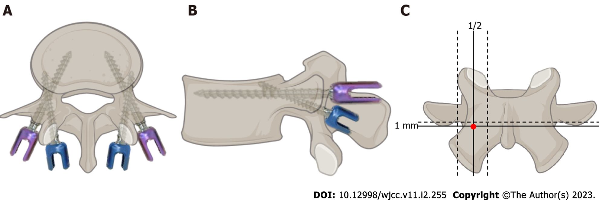
Figure 1 Schematic demonstrating cortical bone trajectory screws (purple) and pedicle screws (blue).
A: Axial view; B: Lateral view; C: The ideal insertion points of a cortical bone trajectory screw (red dot).
- Citation: Peng SB, Yuan XC, Lu WZ, Yu KX. Application of the cortical bone trajectory technique in posterior lumbar fixation. World J Clin Cases 2023; 11(2): 255-267
- URL: https://www.wjgnet.com/2307-8960/full/v11/i2/255.htm
- DOI: https://dx.doi.org/10.12998/wjcc.v11.i2.255


