Copyright
©The Author(s) 2023.
World J Clin Cases. Jul 6, 2023; 11(19): 4723-4728
Published online Jul 6, 2023. doi: 10.12998/wjcc.v11.i19.4723
Published online Jul 6, 2023. doi: 10.12998/wjcc.v11.i19.4723
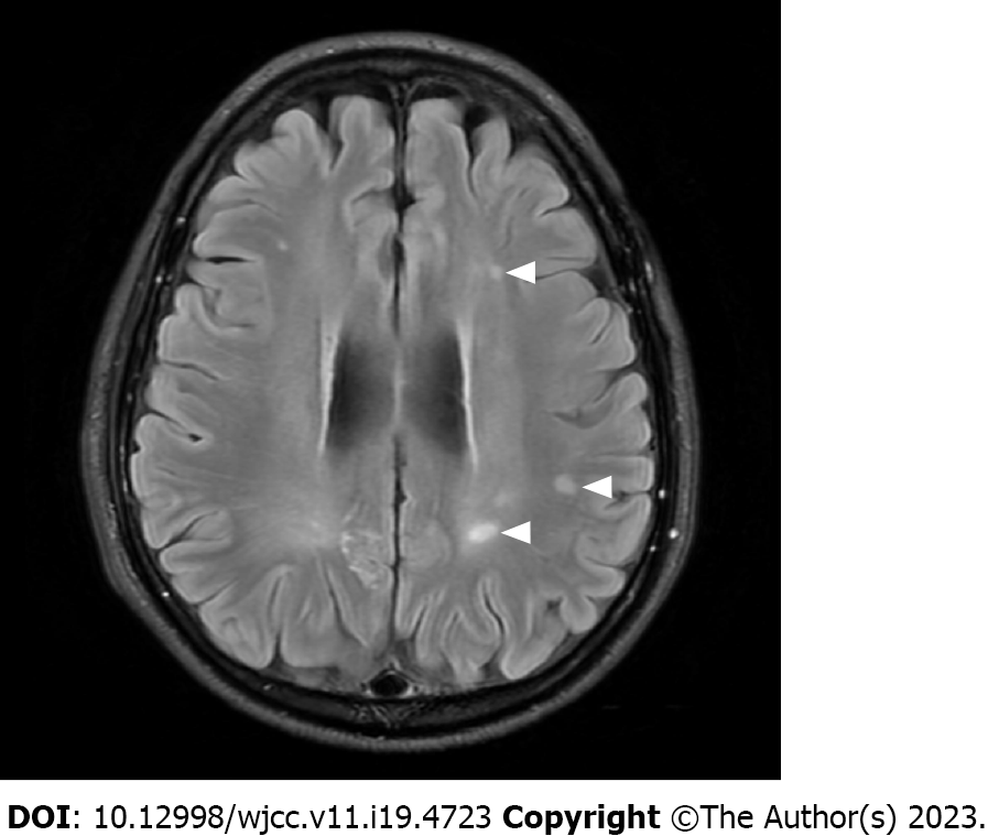
Figure 3 Follow-up magnetic resonance imaging.
Fluid attenuation inversion recovery image performed at one year after clipping surgery shows that several lesions (arrow heads) in the subcortical white matter persisted, despite being reduced in size.
- Citation: Hwang J, Cho WH, Cha SH, Ko JK. Posterior reversible encephalopathy syndrome following uneventful clipping of an unruptured intracranial aneurysm: A case report. World J Clin Cases 2023; 11(19): 4723-4728
- URL: https://www.wjgnet.com/2307-8960/full/v11/i19/4723.htm
- DOI: https://dx.doi.org/10.12998/wjcc.v11.i19.4723


