Copyright
©The Author(s) 2023.
World J Clin Cases. Jul 6, 2023; 11(19): 4723-4728
Published online Jul 6, 2023. doi: 10.12998/wjcc.v11.i19.4723
Published online Jul 6, 2023. doi: 10.12998/wjcc.v11.i19.4723
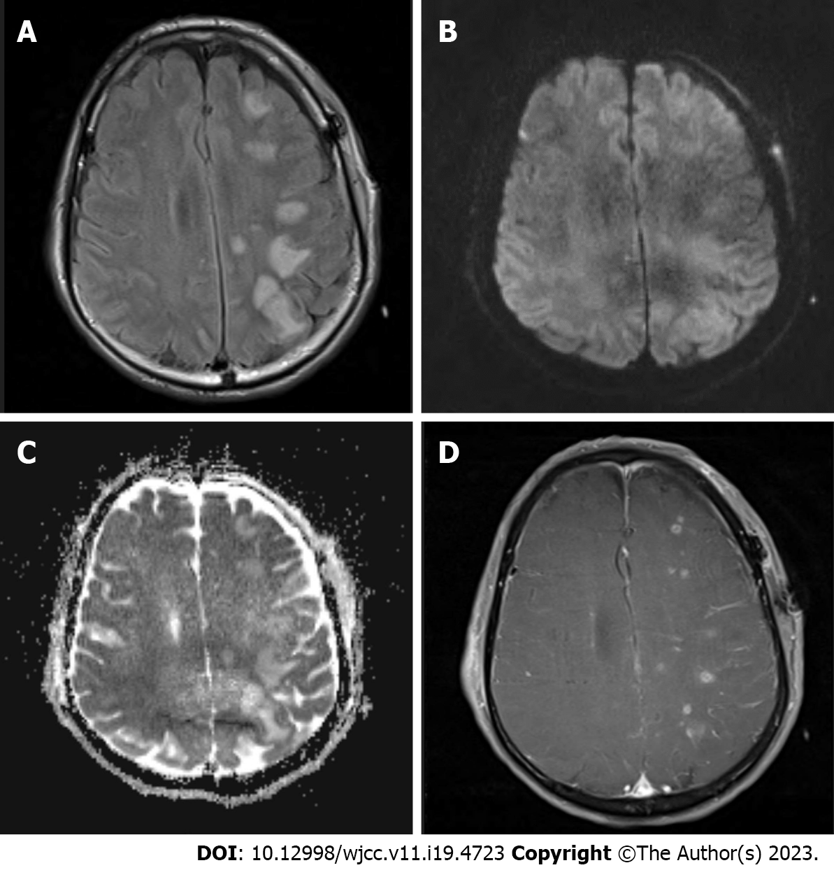
Figure 2 Magnetic resonance imaging obtained immediately following deterioration of consciousness A: Axial fluid attenuation inversion recovery image shows extensive hyperintense lesions predominantly in the cortex and subcortical white matter of the left frontoparietal lobe; B: Diffusion-weighted image shows that lesions are iso to hyperintense without any signs of restricted diffusion; C: Apparent diffusion coefficient maps show that lesions are hyperintense, indicating vasogenic edema; D: On the contrast-enhanced three-dimensional T1-weighted image, lesions show patchy enhancement.
- Citation: Hwang J, Cho WH, Cha SH, Ko JK. Posterior reversible encephalopathy syndrome following uneventful clipping of an unruptured intracranial aneurysm: A case report. World J Clin Cases 2023; 11(19): 4723-4728
- URL: https://www.wjgnet.com/2307-8960/full/v11/i19/4723.htm
- DOI: https://dx.doi.org/10.12998/wjcc.v11.i19.4723


