Copyright
©The Author(s) 2023.
World J Clin Cases. Jul 6, 2023; 11(19): 4707-4712
Published online Jul 6, 2023. doi: 10.12998/wjcc.v11.i19.4707
Published online Jul 6, 2023. doi: 10.12998/wjcc.v11.i19.4707
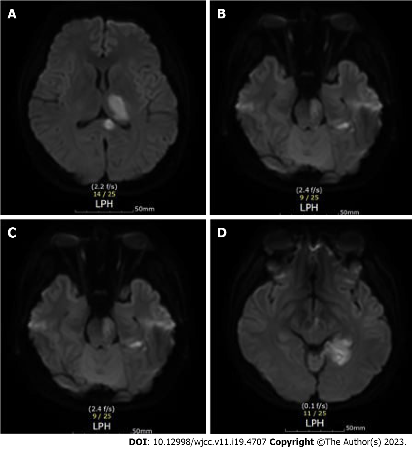
Figure 1 Cerebral infarction lesion shown on a brain magnetic resonance imaging image.
A: Left corpus callosum and thalamus; B: Left occipital lobe; C: Left pons; D: Left midbrain.
- Citation: Jo J, Kim H. Poststroke rehabilitation using repetitive transcranial magnetic stimulation during pregnancy: A case report. World J Clin Cases 2023; 11(19): 4707-4712
- URL: https://www.wjgnet.com/2307-8960/full/v11/i19/4707.htm
- DOI: https://dx.doi.org/10.12998/wjcc.v11.i19.4707


