Copyright
©The Author(s) 2023.
World J Clin Cases. Jul 6, 2023; 11(19): 4579-4600
Published online Jul 6, 2023. doi: 10.12998/wjcc.v11.i19.4579
Published online Jul 6, 2023. doi: 10.12998/wjcc.v11.i19.4579
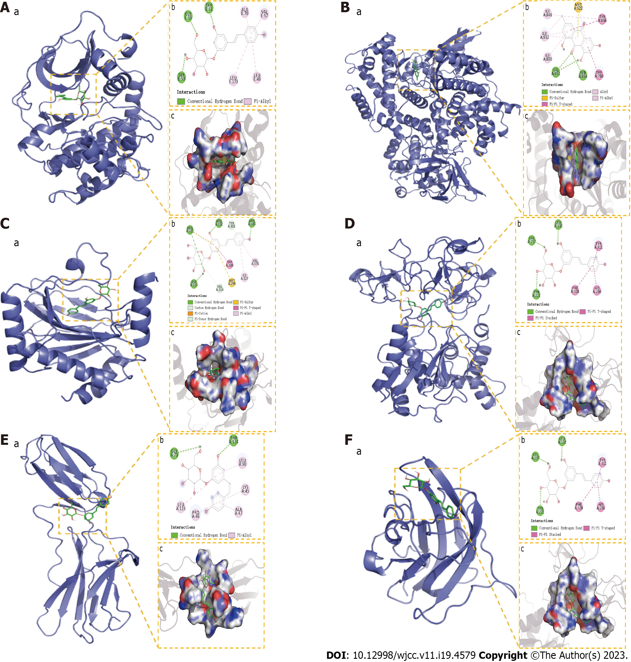
Figure 13 Molecular docking results.
A: The binding mode of RAC-alpha serine/threonine-protein kinase with Polydatin; B: The binding mode of Phosphoinositide 3-Kinase with Emodin; C: The binding mode of hypoxia-inducible factor-1a with Polydatin; D: The binding mode of matrix metalloproteinase-9 with Polydatin; E: The binding mode of IL6 with Polydatin; F: The binding mode of VEGFA with Polydatin. “a” is the 3D structure of the complex, “b” shows the hydrogen bond donor receptor network of the complex, “c” is the 2D binding mode of the complex. AKT: RAC-alpha serine/threonine-protein kinase; PI3K: Phosphoinositide 3-Kinase; HIF1a: hypoxia-inducible factor-1a; MMP9: Matrix metalloproteinase-9; IL6: Interleukin-6; VEGFA: Vascular endothelial growth factor A.
- Citation: Zheng JL, Wang X, Song Z, Zhou P, Zhang GJ, Diao JJ, Han CE, Jia GY, Zhou X, Zhang BQ. Network pharmacology and molecular docking to explore Polygoni Cuspidati Rhizoma et Radix treatment for acute lung injury. World J Clin Cases 2023; 11(19): 4579-4600
- URL: https://www.wjgnet.com/2307-8960/full/v11/i19/4579.htm
- DOI: https://dx.doi.org/10.12998/wjcc.v11.i19.4579


