Copyright
©The Author(s) 2023.
World J Clin Cases. Jul 6, 2023; 11(19): 4477-4497
Published online Jul 6, 2023. doi: 10.12998/wjcc.v11.i19.4477
Published online Jul 6, 2023. doi: 10.12998/wjcc.v11.i19.4477
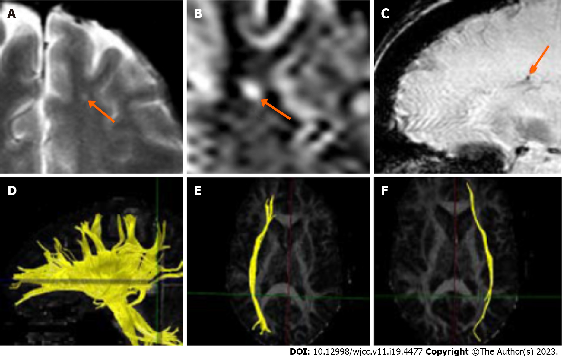
Figure 1 Imaging evidence of a traumatic brain injury in a representative patient.
A: Bright spot on T2-weighted images near the gray-white junction, consistent with traumatic axonal injury (orange arrow); B: Close-up of lesion shown in Panel A (orange arrow); C: Microhemorrhage near the gray-white junction shown on a microhemorrhage-sensitive magnetic resonance image (orange arrow); D: Gaps in the normally continuous distribution of tracts that pass through the corpus callosum; E: Diffusion tensor imaging (DTI) tractography of the relatively normal right fronto-occipital fasciculus; F: DTI tractography of the left fronto-occipital fasciculus showing decreased tracts compared to the right side shown in Panel E.
- Citation: van Velkinburgh JC, Herbst MD, Casper SM. Diffusion tensor imaging in the courtroom: Distinction between scientific specificity and legally admissible evidence. World J Clin Cases 2023; 11(19): 4477-4497
- URL: https://www.wjgnet.com/2307-8960/full/v11/i19/4477.htm
- DOI: https://dx.doi.org/10.12998/wjcc.v11.i19.4477


