Copyright
©The Author(s) 2023.
World J Clin Cases. Jun 26, 2023; 11(18): 4397-4405
Published online Jun 26, 2023. doi: 10.12998/wjcc.v11.i18.4397
Published online Jun 26, 2023. doi: 10.12998/wjcc.v11.i18.4397
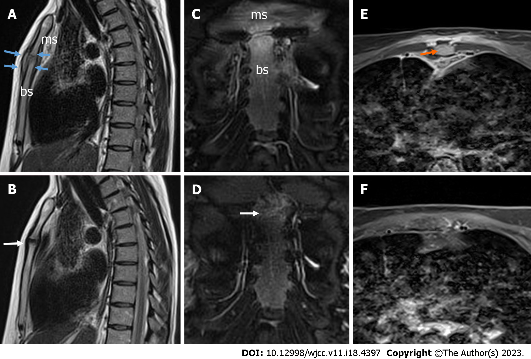
Figure 3 Magnetic resonance imaging of the septic manubriosternal joint.
A: Magnetic resonance imaging (MRI) sagittal T2-weighted blade image. The surfaces of the manubriosternal (MS) joint are eroded in the margins, the joint is enlarged, and the pseudo-capsule is thickened (blue arrows), with visible arthritis; B: MRI sagittal T2-weighted blade image. Four-month follow-up results - the surfaces of the MS joint are further eroded in the margins, perichondritis (white arrow). The pseudo-capsule is thinned; C: MRI coronal T2-weighted blade fat-suppressed image. A mild bone marrow edema is detected; D: MRI coronal T2-weighted blade fat-suppressed image. Four-month follow-up results - a very mild bone marrow edema was detected (white arrow); E: MRI axial contrast-enhanced T1-weighted Dixon water-only image. Localized fluid collection (abscess) up to 12 mm × 4 mm × 44 mm in front of the left first rib was detected without any sign of mediastinitis (orange arrow); F: MRI axial contrast-enhanced T1-weighted Dixon water-only image. Abscesses were not detected; ms: Manubrium of the sternum; bs: Body of the sternum.
- Citation: Seskute G, Kausaite D, Chalkovskaja A, Bulotaite E, Butrimiene I. Diagnostic use of superb microvascular imaging in evaluating septic arthritis of the manubriosternal joint: A case report. World J Clin Cases 2023; 11(18): 4397-4405
- URL: https://www.wjgnet.com/2307-8960/full/v11/i18/4397.htm
- DOI: https://dx.doi.org/10.12998/wjcc.v11.i18.4397


