Copyright
©The Author(s) 2023.
World J Clin Cases. Jun 26, 2023; 11(18): 4384-4391
Published online Jun 26, 2023. doi: 10.12998/wjcc.v11.i18.4384
Published online Jun 26, 2023. doi: 10.12998/wjcc.v11.i18.4384
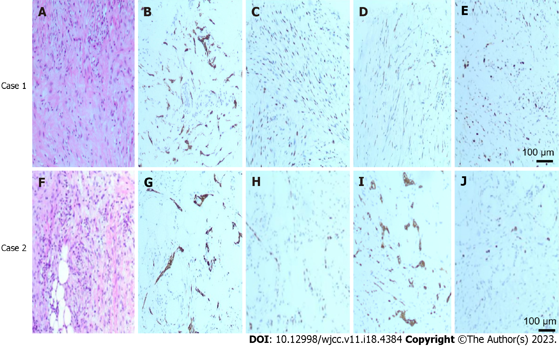
Figure 2 Puncture pathology and immunohistochemistry of right breast mass.
A: HE staining showed that the tumor was composed of spindle cells with mild morphology and mild atypia of the tumor cells; B: Tumor cells were positive for CKPan by Envision assay; C: Envision test showed that tumor cells were positive for p63; D: Envision test showed tumor cells positive for CK5/6; E: Envision test showed approximately 15% Ki-67 for tumor cells. F: HE stained tumor was composed of spindle cells with mild morphology and mild atypia of tumor cells; G: Tumor cells were positive for CKPan by Envision assay; H: Tumor cells were positive for p63 by Envision assay; I: Tumor cells were partially positive for CK5/6 by Envision assay; J: Ki-67 hotspot area of tumor cells detected by Envision method was about + 25%. Original magnification: × 400; Scale bars: 100 μm.
- Citation: Bao WY, Zhou JH, Luo Y, Lu Y. Fibromatosis-like metaplastic carcinoma of the breast: Two case reports. World J Clin Cases 2023; 11(18): 4384-4391
- URL: https://www.wjgnet.com/2307-8960/full/v11/i18/4384.htm
- DOI: https://dx.doi.org/10.12998/wjcc.v11.i18.4384


