Copyright
©The Author(s) 2023.
World J Clin Cases. Jun 26, 2023; 11(18): 4377-4383
Published online Jun 26, 2023. doi: 10.12998/wjcc.v11.i18.4377
Published online Jun 26, 2023. doi: 10.12998/wjcc.v11.i18.4377
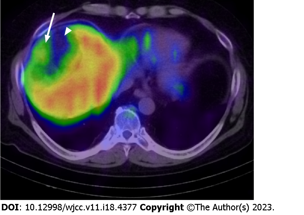
Figure 5 A fusion image with single photon emission computed tomography using 111InCl3 and computed tomography.
The image shows the mild accumulation of radiopharmaceutical in the peripheral area of the lesion (white arrow). Poor accumulation was observed in the central region corresponding to the dominant fat area (white arrowhead).
- Citation: Sato A, Saito K, Abe K, Sugimoto K, Nagao T, Sukeda A, Yunaiyama D. Indium chloride bone marrow scintigraphy for hepatic myelolipoma: A case report. World J Clin Cases 2023; 11(18): 4377-4383
- URL: https://www.wjgnet.com/2307-8960/full/v11/i18/4377.htm
- DOI: https://dx.doi.org/10.12998/wjcc.v11.i18.4377


