Copyright
©The Author(s) 2023.
World J Clin Cases. Jun 26, 2023; 11(18): 4377-4383
Published online Jun 26, 2023. doi: 10.12998/wjcc.v11.i18.4377
Published online Jun 26, 2023. doi: 10.12998/wjcc.v11.i18.4377
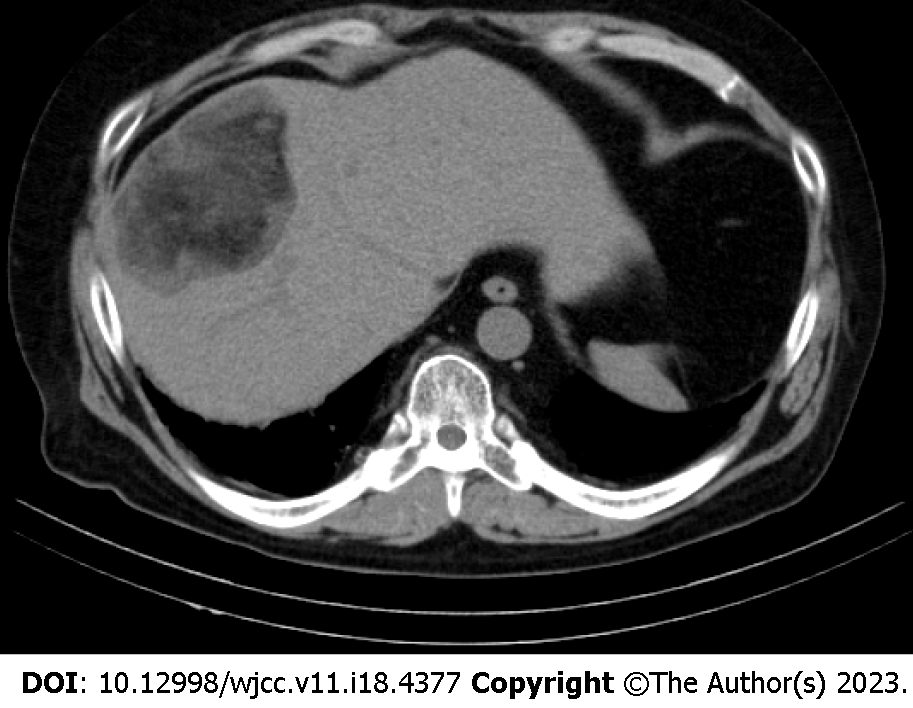
Figure 2 Plain computed tomography.
The image shows a well-defined lesion in segment 8 of the liver. The center of the lesion mainly shows the fat density and the peripheral area of the lesion shows hypodensity.
- Citation: Sato A, Saito K, Abe K, Sugimoto K, Nagao T, Sukeda A, Yunaiyama D. Indium chloride bone marrow scintigraphy for hepatic myelolipoma: A case report. World J Clin Cases 2023; 11(18): 4377-4383
- URL: https://www.wjgnet.com/2307-8960/full/v11/i18/4377.htm
- DOI: https://dx.doi.org/10.12998/wjcc.v11.i18.4377


