Copyright
©The Author(s) 2023.
World J Clin Cases. Jun 26, 2023; 11(18): 4341-4349
Published online Jun 26, 2023. doi: 10.12998/wjcc.v11.i18.4341
Published online Jun 26, 2023. doi: 10.12998/wjcc.v11.i18.4341
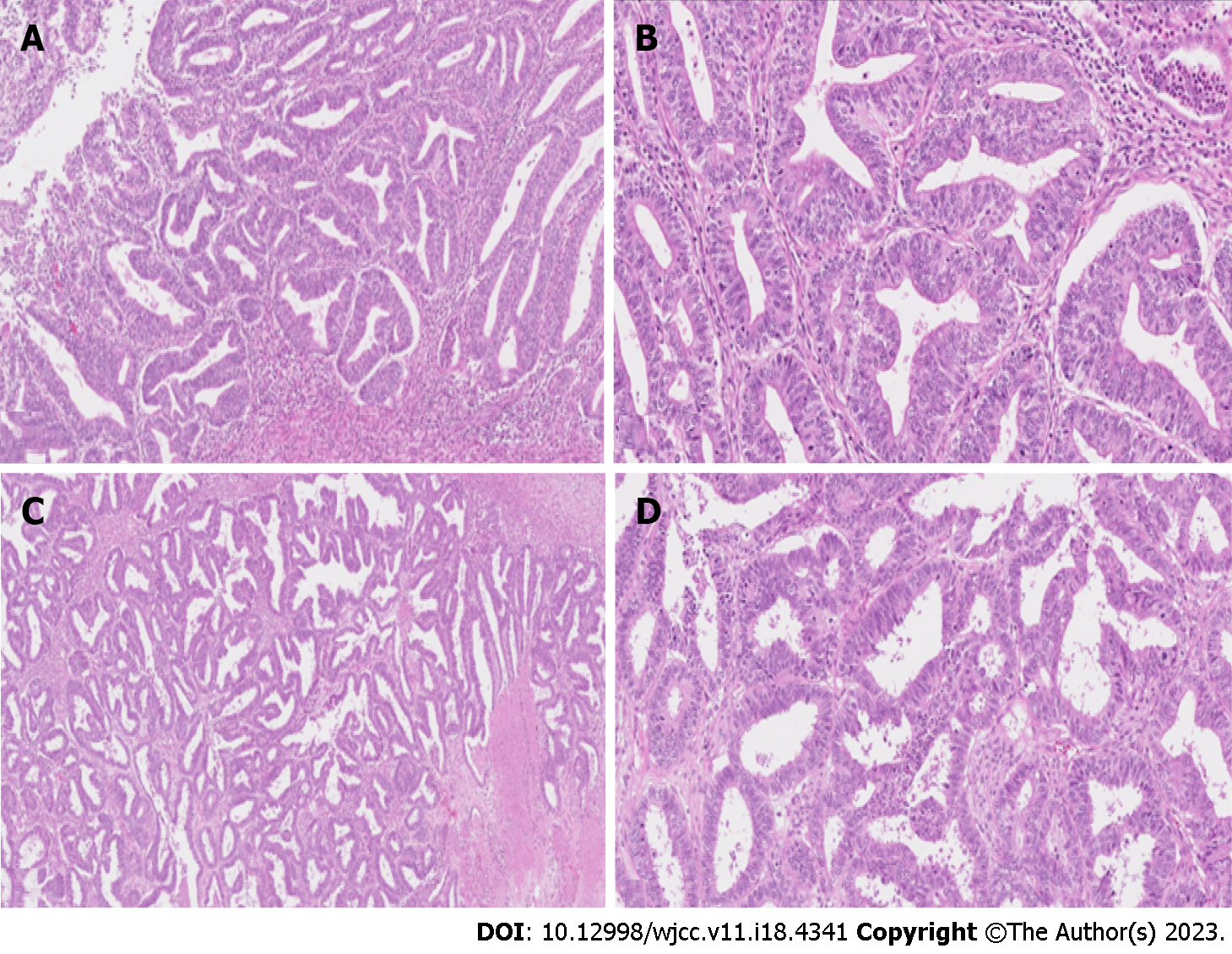
Figure 2 Haematoxylin and eosin staining in case II.
A: Haematoxylin and eosin-stained (H&E) slide at low power (lens objective × 4) shows closely packed irregular glandular structures; B: H&E-stained slide at medium power (lens objective × 14) shows tall columnar epithelium with oval, stratified nuclei with visible nucleoli; C: H&E-stained slide at low power (lens objective × 4) shows closely packed irregular glandular structures with focal necrosis; D: H&E-stained slide at medium power (lens objective × 14) shows columnar epithelium with oval, stratified nuclei, and visible nucleoli.
- Citation: Žilovič D, Čiurlienė R, Šidlovska E, Vaicekauskaitė I, Sabaliauskaitė R, Jarmalaitė S. Synchronous endometrial and ovarian cancer: A case report. World J Clin Cases 2023; 11(18): 4341-4349
- URL: https://www.wjgnet.com/2307-8960/full/v11/i18/4341.htm
- DOI: https://dx.doi.org/10.12998/wjcc.v11.i18.4341


