Copyright
©The Author(s) 2023.
World J Clin Cases. Jun 26, 2023; 11(18): 4334-4340
Published online Jun 26, 2023. doi: 10.12998/wjcc.v11.i18.4334
Published online Jun 26, 2023. doi: 10.12998/wjcc.v11.i18.4334
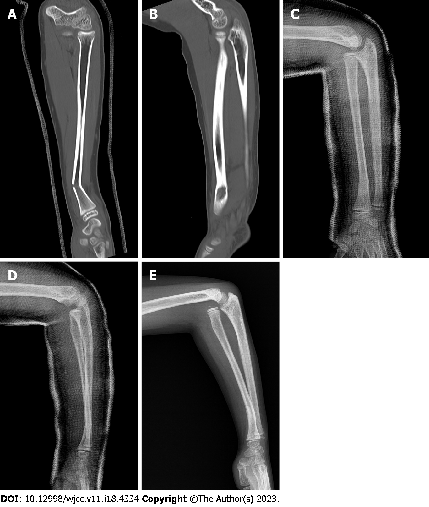
Figure 1 Images.
A and B: Computed tomography images of the left forearm performed at the time of injury. A linear fracture with angulation is seen in the distal third of the radius (A); there is no radial head dislocation in the elbow joint (B); C and D: Radiographs of the left forearm performed two weeks after the injury. The radial fracture is well reduced and shows no evidence of displacement progression (C); a mild subluxation of the radial head, which was not observed at the time of injury, is seen at the elbow joint (D); E: Preoperative radiographs of the left forearm performed three months after the injury. Malunion with a posterior convex deformity of 22° at the radial fracture site associated with complete anterior dislocation of the radial head is shown.
- Citation: Kim KB, Wang SI. Delayed dislocation of the radial head associated with malunion of distal radial fracture: A case report. World J Clin Cases 2023; 11(18): 4334-4340
- URL: https://www.wjgnet.com/2307-8960/full/v11/i18/4334.htm
- DOI: https://dx.doi.org/10.12998/wjcc.v11.i18.4334


