Copyright
©The Author(s) 2023.
World J Clin Cases. Jun 26, 2023; 11(18): 4295-4305
Published online Jun 26, 2023. doi: 10.12998/wjcc.v11.i18.4295
Published online Jun 26, 2023. doi: 10.12998/wjcc.v11.i18.4295
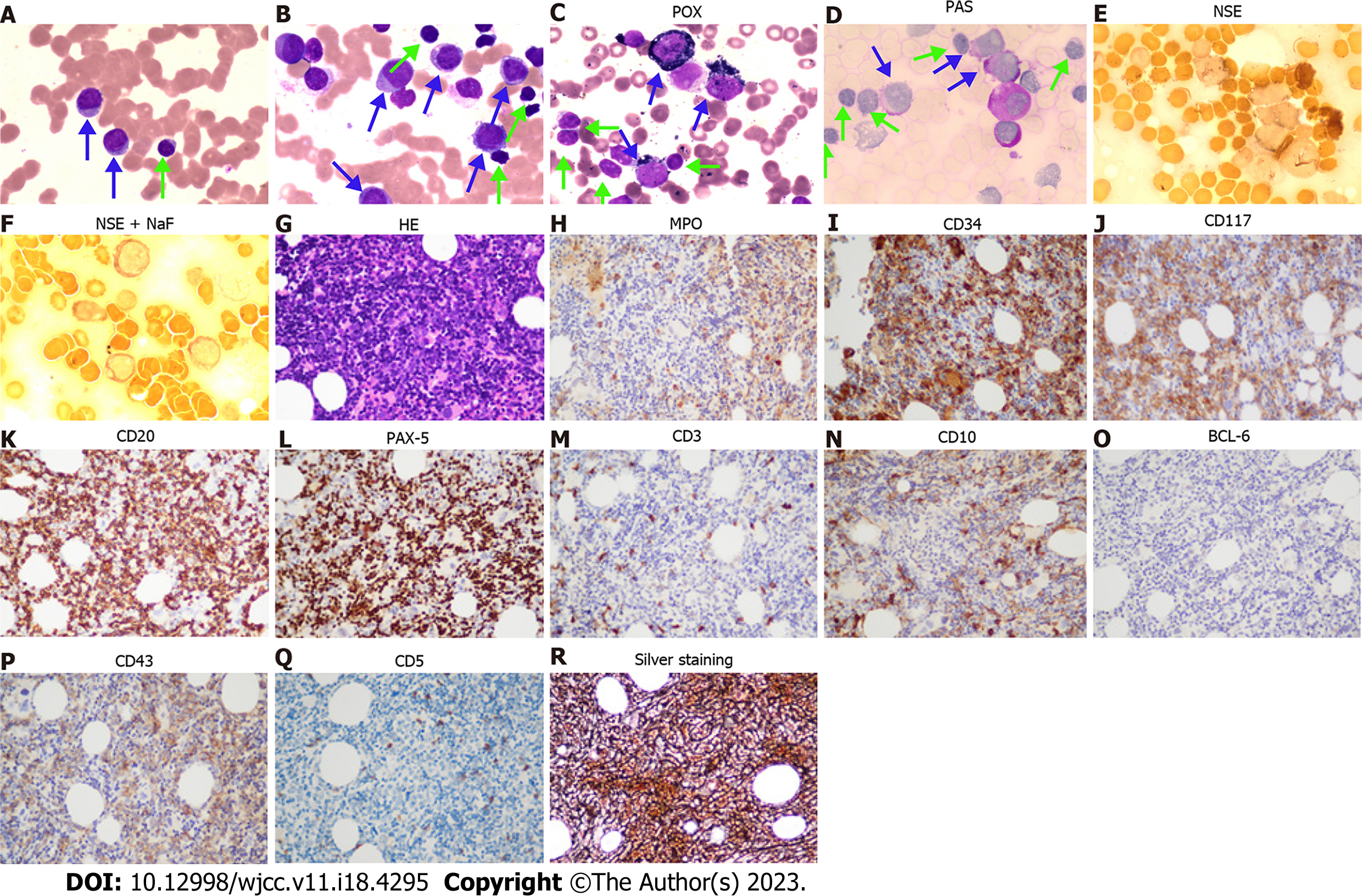
Figure 2 The cytomorphology of peripheral blood and bone marrow.
A: Peripheral blood smear (Wright staining, 100 × objective); B: Bone marrow smear (Wright staining, 100 × objective); C-F: Histochemistry staining for bone marrow smear. Peroxidase (52%, 99 points), periodic acid-Schiff stain (86%, 158 points), non-specific esterase (NSE) (32%, 35 points), and NSE + NaF (25%, 26 points) staining for bone marrow smear (Blue arrows direct myeloid blasts, while green arrows direct small lymphocytes); G-R: Bone marrow biopsy (40 × objective); G: Hematoxylin-Eosin staining; H-R: Immunohistochemistry shows myeloperoxidase (myelocytes +), CD34 (myelocytes ++), CD117 (myelocytes +), CD20 (B cells ++), PAX-5 (B cells ++), CD3 (T cells, few +), CD10 (few +), BCL-6(-), CD43(partial +), CD5 (T cells -), and silver staining (+++). POX: Peroxidase; PAS: Periodic acid-Schiff; NSE: Non-specific esterase; MPO: Myeloperoxidase.
- Citation: Zhang LB, Zhang L, Xin HL, Wang Y, Bao HY, Meng QQ, Jiang SY, Han X, Chen WR, Wang JN, Shi XF. Coexistence of diffuse large B-cell lymphoma, acute myeloid leukemia, and untreated lymphoplasmacytic lymphoma/waldenström macroglobulinemia in a same patient: A case report. World J Clin Cases 2023; 11(18): 4295-4305
- URL: https://www.wjgnet.com/2307-8960/full/v11/i18/4295.htm
- DOI: https://dx.doi.org/10.12998/wjcc.v11.i18.4295


