Copyright
©The Author(s) 2023.
World J Clin Cases. Jun 26, 2023; 11(18): 4251-4257
Published online Jun 26, 2023. doi: 10.12998/wjcc.v11.i18.4251
Published online Jun 26, 2023. doi: 10.12998/wjcc.v11.i18.4251
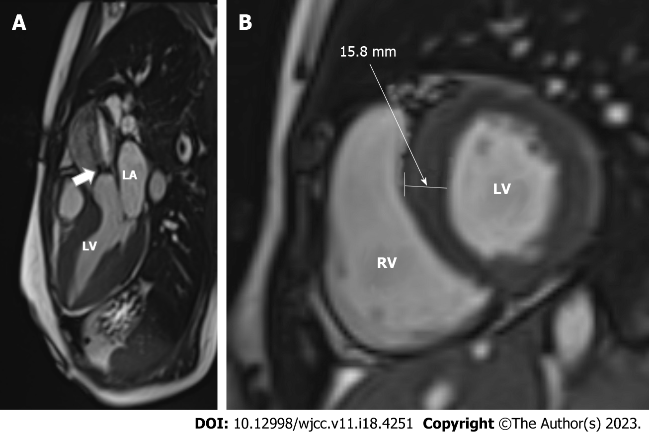
Figure 3 Cardiac magnetic resonance imaging was acquired on a 1.
5T scanner. A: Systolic jet of aortic stenosis (arrow) can be observed on a 3-chamber view cine steady-state free precession image; B: The left ventricular myocardium was hypertrophic with a 15.8-mm-thick interventricular septum on a short-axis SSFP enddiastolic image. LA: Left atrium; LV: Left ventricle; RV: Right ventricle; LV: Left ventricle.
- Citation: Sopek Merkaš I, Lakušić N, Predrijevac M, Štambuk K, Hrabak Paar M. Bicuspid aortic valve with associated aortopathy, significant left ventricular hypertrophy or concomitant hypertrophic cardiomyopathy: A diagnostic and therapeutic challenge. World J Clin Cases 2023; 11(18): 4251-4257
- URL: https://www.wjgnet.com/2307-8960/full/v11/i18/4251.htm
- DOI: https://dx.doi.org/10.12998/wjcc.v11.i18.4251


