Copyright
©The Author(s) 2023.
World J Clin Cases. Jun 16, 2023; 11(17): 4168-4178
Published online Jun 16, 2023. doi: 10.12998/wjcc.v11.i17.4168
Published online Jun 16, 2023. doi: 10.12998/wjcc.v11.i17.4168
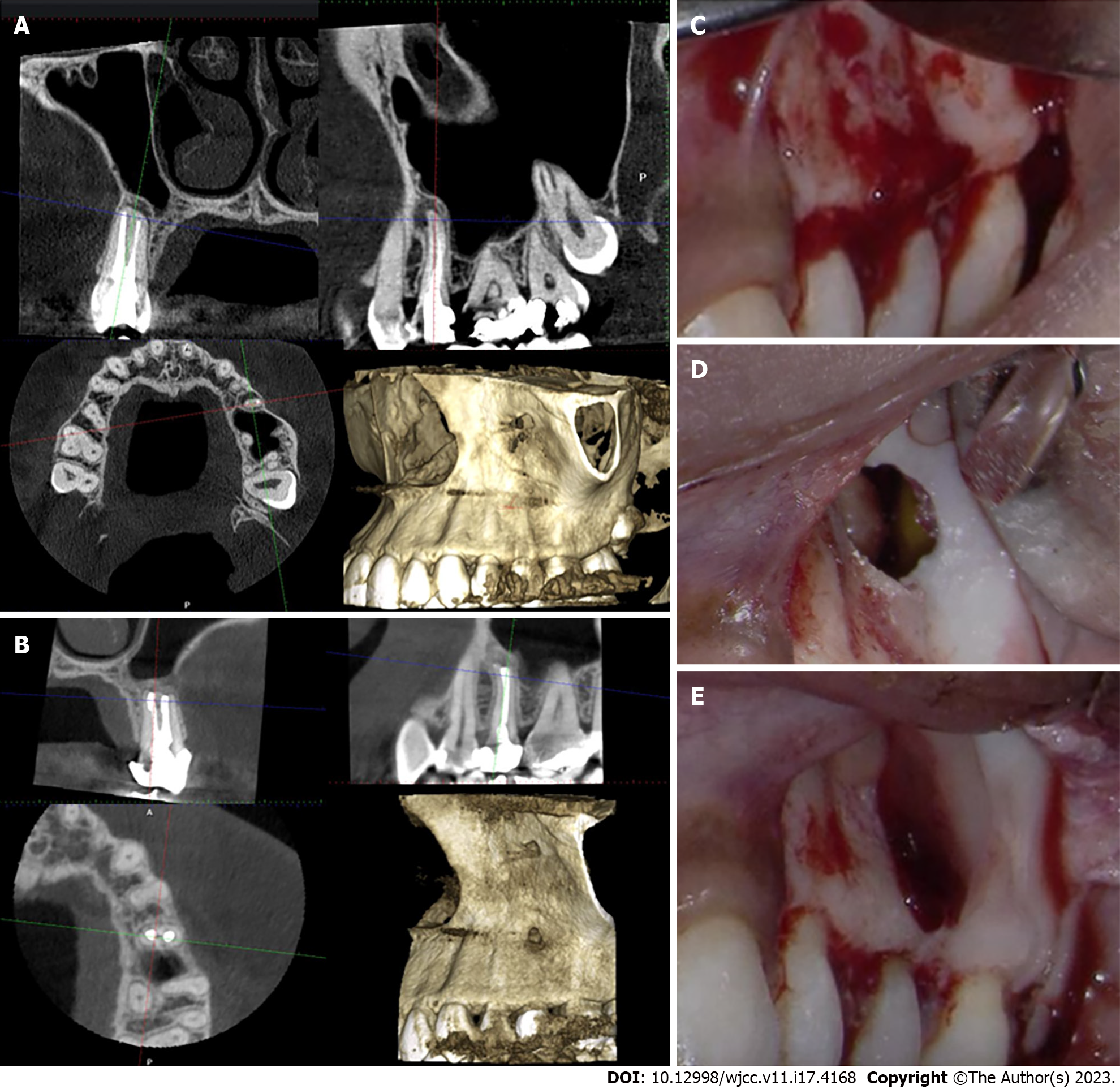
Figure 2 Periapical endodontic surgery on the maxillary left second premolar using advanced platelet-rich fibrin membrane.
A: The cone-beam computed tomography (CBCT) shows localized radiolucency surrounding the apex of the maxillary left second premolar in the sagittal section. There is a thin layer of the labial bone surrounding the apex and a displaced sinus periosteum upward which is a typical feature of periapical osteoperiostitis in the sagittal section. The root canal treatment appears to be adequate with a uniform density; B: The CBCT shows bony healing of the resected area within four months and a normal appearance of the maxillary sinus; C: A clinical photo of the labial bone before root resection; D: A clinical photo of the bony defect after resection shows a hollow space adjacent to the maxillary sinus; E: The platelet-rich fibrin membrane is used to seal the bony defect.
- Citation: Algahtani FN, Almohareb R, Aljamie M, Alkhunaini N, ALHarthi SS, Barakat R. Application of advanced platelet-rich fibrin for through-and-through bony defect during endodontic surgery: Three case reports and review of the literature. World J Clin Cases 2023; 11(17): 4168-4178
- URL: https://www.wjgnet.com/2307-8960/full/v11/i17/4168.htm
- DOI: https://dx.doi.org/10.12998/wjcc.v11.i17.4168


