Copyright
©The Author(s) 2023.
World J Clin Cases. Jun 16, 2023; 11(17): 4152-4158
Published online Jun 16, 2023. doi: 10.12998/wjcc.v11.i17.4152
Published online Jun 16, 2023. doi: 10.12998/wjcc.v11.i17.4152
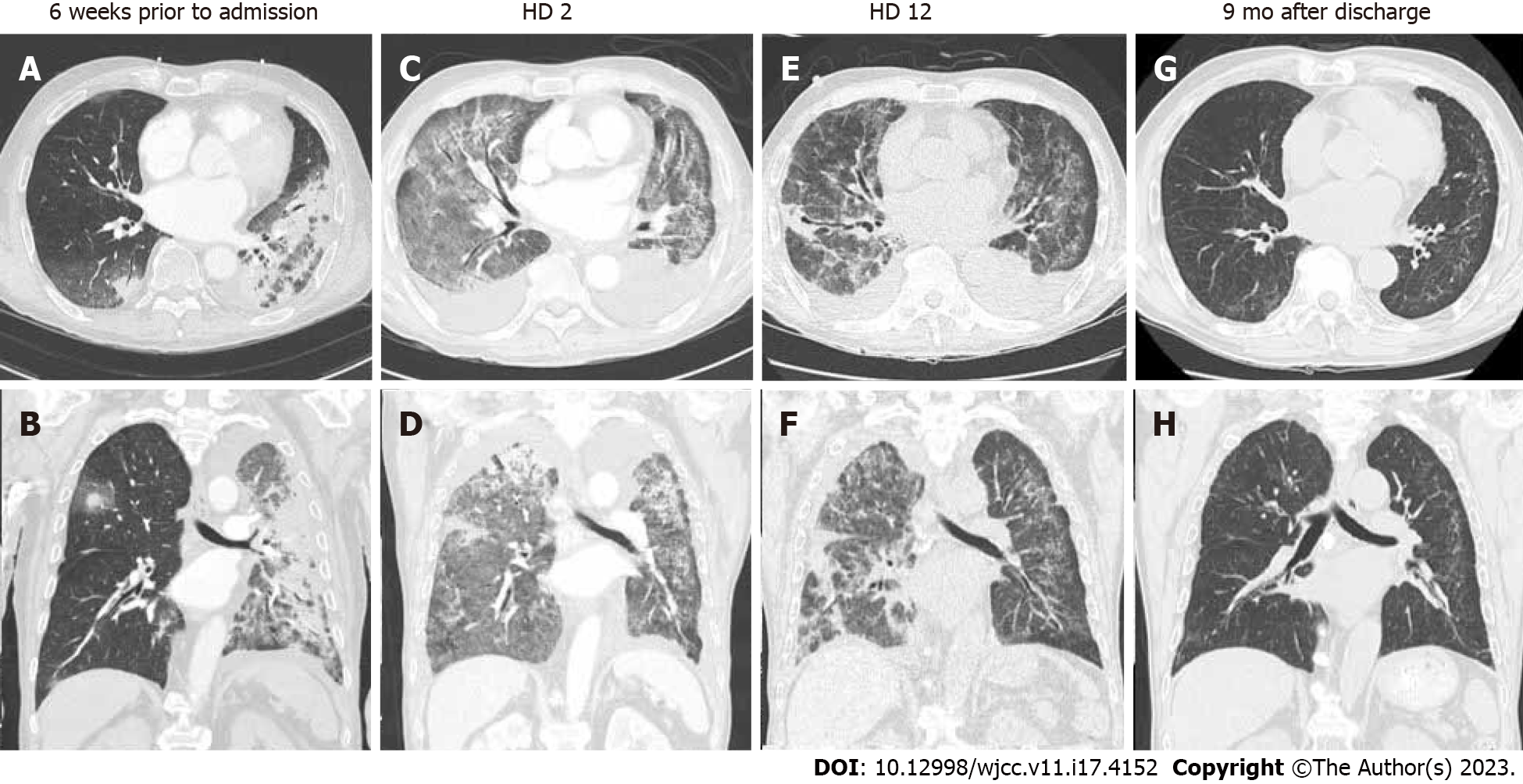
Figure 2 Serial chest computed tomography images in time of coronavirus disease 2019, HD 2, HD 12, and 9 mo after discharge.
A and B: Chest computed tomography (CT) scans performed at the time of coronavirus disease 2019 (COVID-19) (6 wk prior to admission) show peribronchovascular distributed consolidations in both lungs, with greater extent in left lung; this is consistent with COVID-19 pneumonia; C and D: Chest CT scans performed on admission show diffuse ground-glass opacities and patchy consolidations in both lungs and small to moderate amount of bilateral pleural effusions; E and F: Follow-up low-dose chest CT scans after steroid pulse therapy show decreased extent and density of bilateral parenchymal opacifications and more progressed bronchial dilatation; G and H: Follow-up chest CT scans after discharge show almost resolution of bilateral lung disease with mild residual ground-glass opacities in peripheral lungs.
- Citation: Park S, Jang Y, Koo SM, Nam BD, Yoon HY. Glucocorticoid pulse therapy in an elderly patient with post-COVID-19 organizing pneumonia: A case report. World J Clin Cases 2023; 11(17): 4152-4158
- URL: https://www.wjgnet.com/2307-8960/full/v11/i17/4152.htm
- DOI: https://dx.doi.org/10.12998/wjcc.v11.i17.4152


