Copyright
©The Author(s) 2023.
World J Clin Cases. Jun 16, 2023; 11(17): 4105-4116
Published online Jun 16, 2023. doi: 10.12998/wjcc.v11.i17.4105
Published online Jun 16, 2023. doi: 10.12998/wjcc.v11.i17.4105
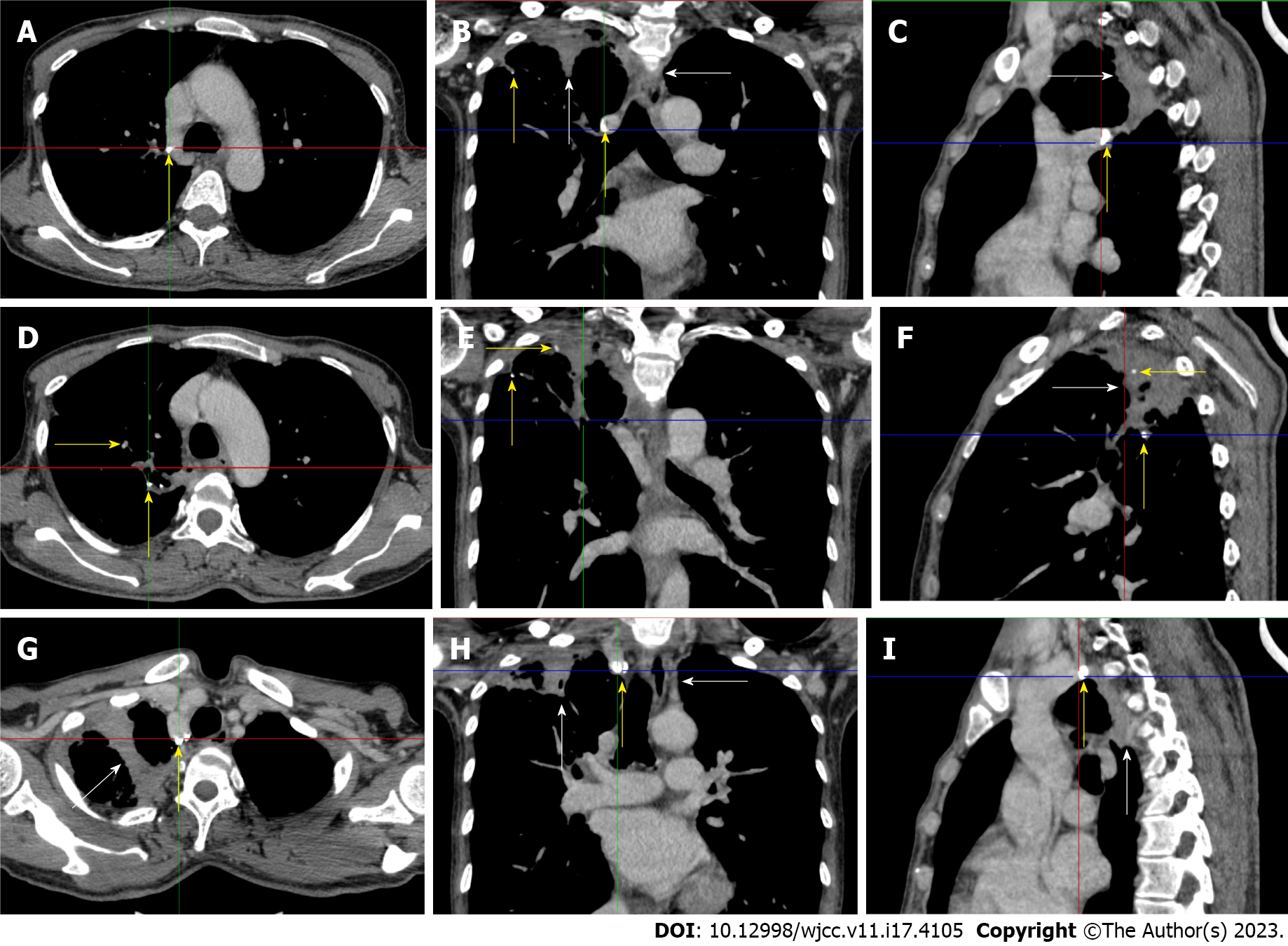
Figure 2 Chest computed tomography scan in the flared inflammatory episode.
Multiple calcified lesions (yellow arrows) with adjacent exudative lesions (white arrows) were present in the right upper lung and mediastinal lymph nodes, indicating the presence of old tuberculosis infection. A-C: A large calcified lesion was present in the posterior mediastinum with adjacent exudative lesions; D-F: Successional exudative lesions on a background of calcified lesions formed a large fused exudative lesion present in the right upper lung abutting the pleura and mediastinum; G-I: A large calcified lesion was present in the top of the right upper lung with adjacent exudative lesions. This imaging feature is typical of the reactivation of an old tuberculosis infection.
- Citation: Ju B, Xiu NN, Xu J, Yang XD, Sun XY, Zhao XC. Flared inflammatory episode transforms advanced myelodysplastic syndrome into aplastic pancytopenia: A case report and literature review. World J Clin Cases 2023; 11(17): 4105-4116
- URL: https://www.wjgnet.com/2307-8960/full/v11/i17/4105.htm
- DOI: https://dx.doi.org/10.12998/wjcc.v11.i17.4105


