Copyright
©The Author(s) 2023.
World J Clin Cases. Jun 6, 2023; 11(16): 3921-3928
Published online Jun 6, 2023. doi: 10.12998/wjcc.v11.i16.3921
Published online Jun 6, 2023. doi: 10.12998/wjcc.v11.i16.3921
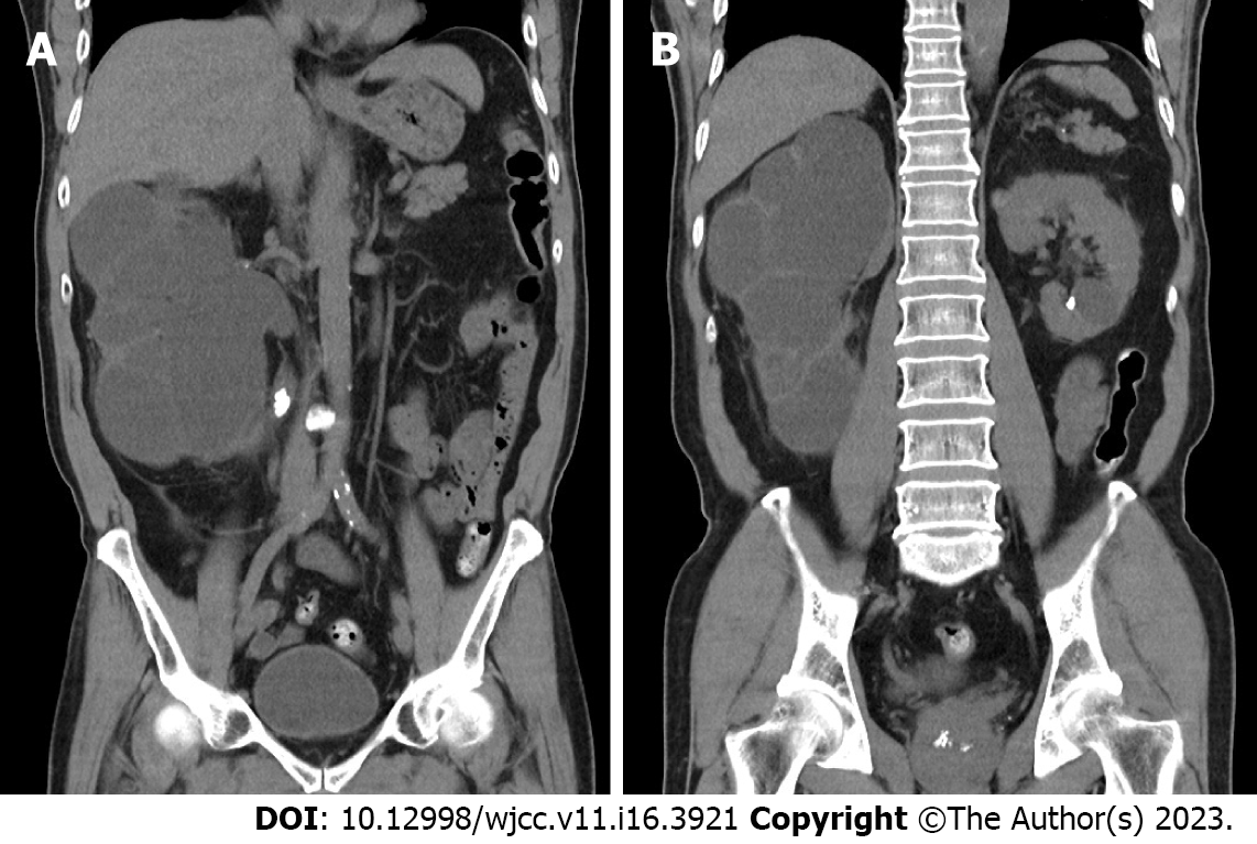
Figure 2 Computed tomography scans.
A: Computed tomography (CT) scans of the abdomen demonstrate severe hydronephrosis of the right kidney with a paper-thin cortex. Upper-third ureteral stone is noted; B: CT scans of the same area revealed a left renal stone with a cyst.
- Citation: Tsai YC, Li CC, Chen BT, Wang CY. Coexistence of urinary tuberculosis and urothelial carcinoma: A case report. World J Clin Cases 2023; 11(16): 3921-3928
- URL: https://www.wjgnet.com/2307-8960/full/v11/i16/3921.htm
- DOI: https://dx.doi.org/10.12998/wjcc.v11.i16.3921


