Copyright
©The Author(s) 2023.
World J Clin Cases. Jun 6, 2023; 11(16): 3915-3920
Published online Jun 6, 2023. doi: 10.12998/wjcc.v11.i16.3915
Published online Jun 6, 2023. doi: 10.12998/wjcc.v11.i16.3915
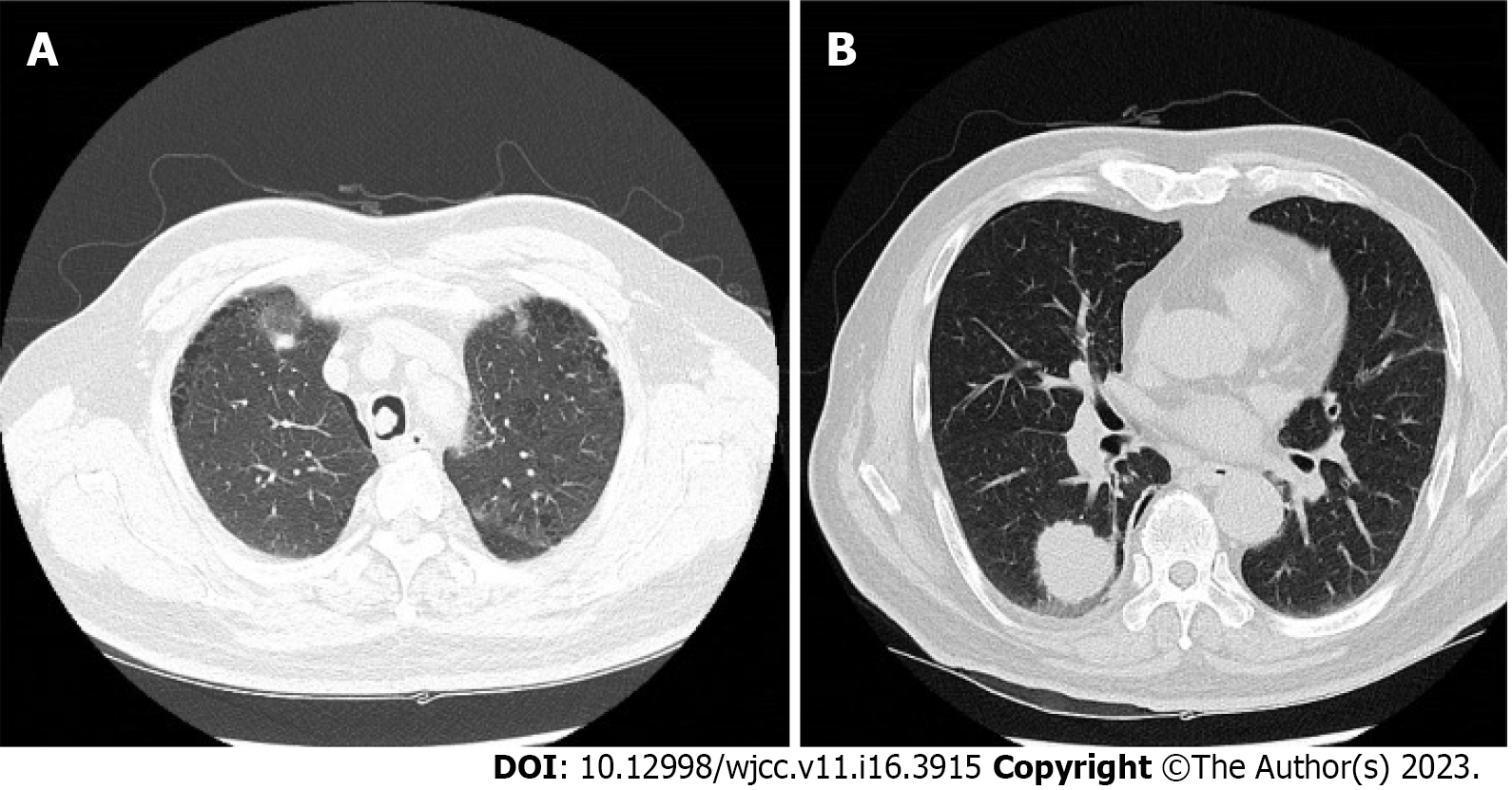
Figure 1 Computed tomography of the patient’s chest.
A: A 10-mm lesion was seen on the right side of the mid-trachea; B: A 30-mm nodule was seen in the right lower lung lobe.
- Citation: Jung HS, Kim HJ, Kim KW. Intraoperative photodynamic therapy for tracheal mass in non-small cell lung cancer: A case report. World J Clin Cases 2023; 11(16): 3915-3920
- URL: https://www.wjgnet.com/2307-8960/full/v11/i16/3915.htm
- DOI: https://dx.doi.org/10.12998/wjcc.v11.i16.3915


