Copyright
©The Author(s) 2023.
World J Clin Cases. Jun 6, 2023; 11(16): 3858-3863
Published online Jun 6, 2023. doi: 10.12998/wjcc.v11.i16.3858
Published online Jun 6, 2023. doi: 10.12998/wjcc.v11.i16.3858
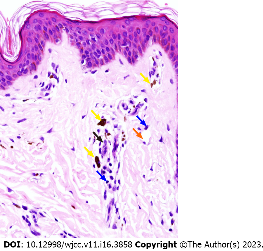
Figure 3 Histopathological findings.
Histopathology showed hyperkeratosis, scattered vacuolar endothelial cells, infiltration of lymphocytes (blue arrows) and histocytes (black arrow) around blood vessels (orange arrow), and deposition of hemosiderin (yellow arrows) in papillary dermis (Hematoxylin eosin staining: Magnification × 400).
- Citation: Pu YJ, Jiang HJ, Zhang L. Purpura annularis telangiectodes of Majocchi: A case report. World J Clin Cases 2023; 11(16): 3858-3863
- URL: https://www.wjgnet.com/2307-8960/full/v11/i16/3858.htm
- DOI: https://dx.doi.org/10.12998/wjcc.v11.i16.3858


