Copyright
©The Author(s) 2023.
World J Clin Cases. Jun 6, 2023; 11(16): 3858-3863
Published online Jun 6, 2023. doi: 10.12998/wjcc.v11.i16.3858
Published online Jun 6, 2023. doi: 10.12998/wjcc.v11.i16.3858
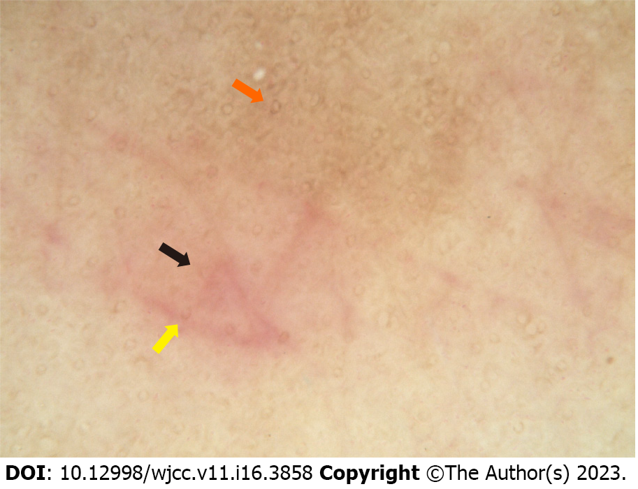
Figure 2 Dermatoscopic appearance of the lesion.
The infiltration method was used (× 50). Dermascopy showed a large number of reticular or honeycomb pigmentation (orange arrow) in the center of the lesion, and lavender patches (black arrow) and a few focally distributed punctate blood vessels (yellow arrow) were seen on the edge of lesion.
- Citation: Pu YJ, Jiang HJ, Zhang L. Purpura annularis telangiectodes of Majocchi: A case report. World J Clin Cases 2023; 11(16): 3858-3863
- URL: https://www.wjgnet.com/2307-8960/full/v11/i16/3858.htm
- DOI: https://dx.doi.org/10.12998/wjcc.v11.i16.3858


