Copyright
©The Author(s) 2023.
World J Clin Cases. May 26, 2023; 11(15): 3571-3577
Published online May 26, 2023. doi: 10.12998/wjcc.v11.i15.3571
Published online May 26, 2023. doi: 10.12998/wjcc.v11.i15.3571
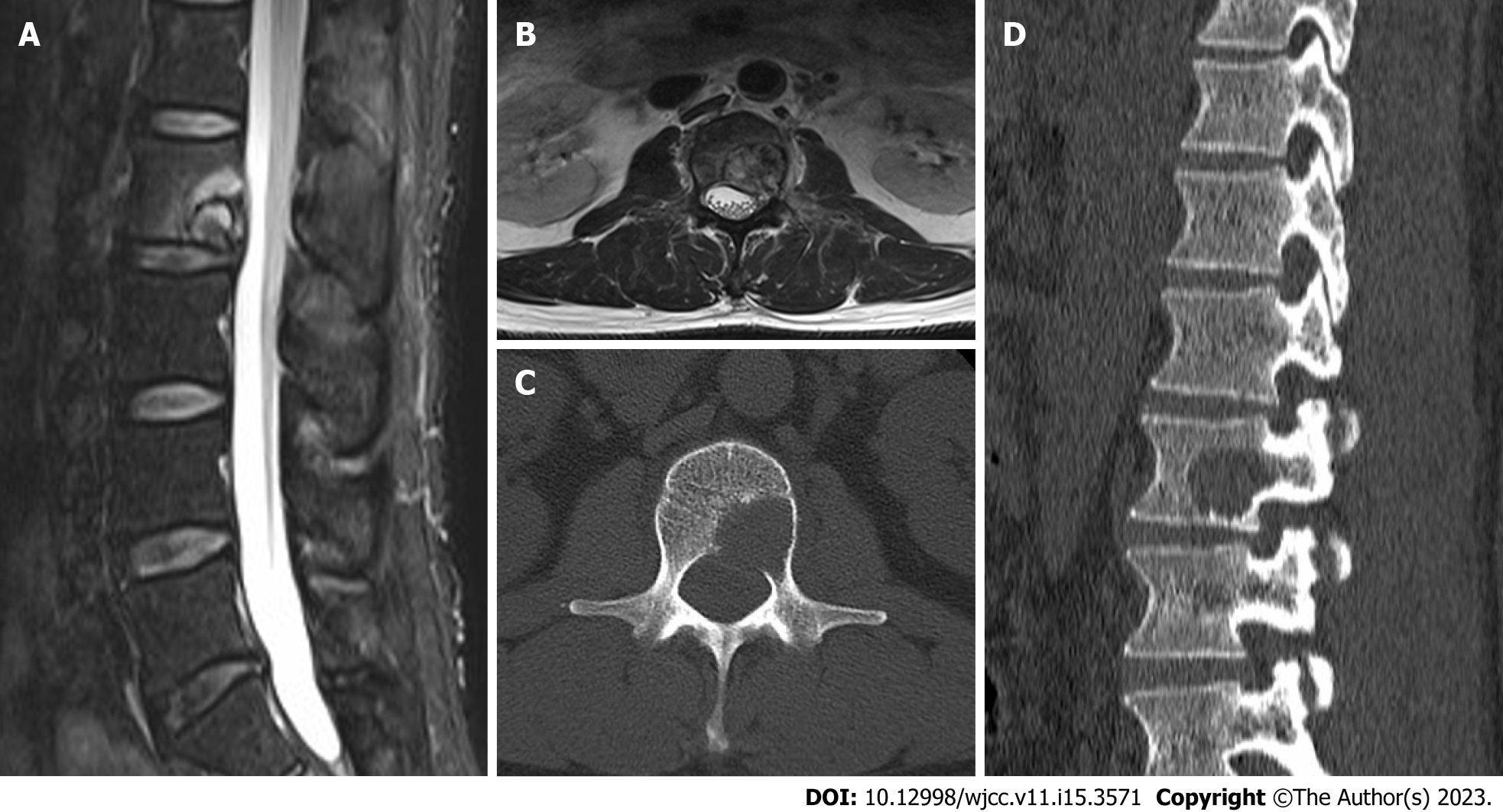
Figure 1 Magnetic resonance imaging and computed tomography of the patient's lumbar spine.
A: In the sagittal position, tumors may be compressing the spinal cord; B: The transverse view shows that the right spinal cord is compressed by a tumor; C: The transverse position shows destruction of the right pedicle and vertebral body; D: In the sagittal position, the posterior side of the spine is destroyed by tumors.
- Citation: Guo ZX, Zhao XL, Zhao ZY, Zhu QY, Wang ZY, Xu M. Malignant melanoma resection and reconstruction with the first manifestation of lumbar metastasis: A case report. World J Clin Cases 2023; 11(15): 3571-3577
- URL: https://www.wjgnet.com/2307-8960/full/v11/i15/3571.htm
- DOI: https://dx.doi.org/10.12998/wjcc.v11.i15.3571


