Copyright
©The Author(s) 2023.
World J Clin Cases. May 26, 2023; 11(15): 3552-3559
Published online May 26, 2023. doi: 10.12998/wjcc.v11.i15.3552
Published online May 26, 2023. doi: 10.12998/wjcc.v11.i15.3552
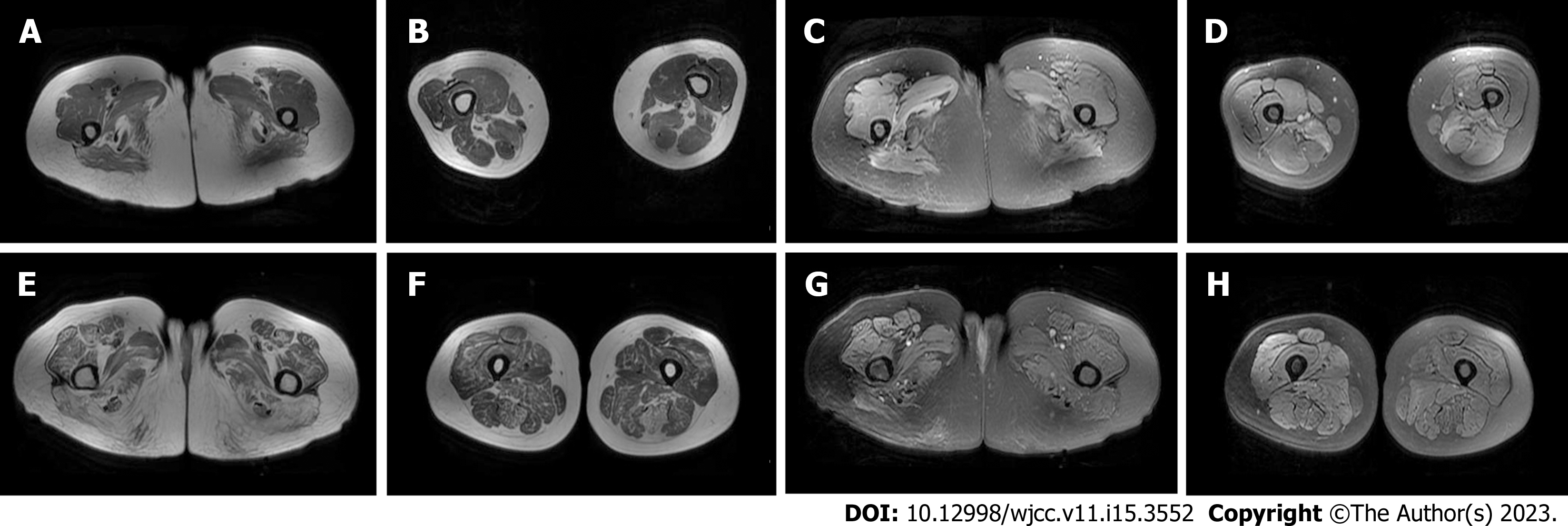
Figure 2 Magnetic resonance imaging of the thigh muscle of patients.
A-D: Patient 1; A and B: T1WI sequence showed that fat infiltration was mainly in the medial and posterior thigh muscle groups; C and D: Short time of inversion recovery (STIR) sequence showed that oedema was mainly in the posterior thigh muscle groups; E-H: Patient 2; E and F: T1WI sequence showed different degrees of fat infiltration in the muscles of the lower extremities, which was more obvious in the posterior group, and the gracilis muscle was relatively preserved; G and H: STIR sequence showed that oedema was found in the right lower limb, mainly concentrated in the anterior external and posterior thigh muscle groups.
- Citation: Chen BH, Zhu XM, Xie L, Hu HQ. Immune-mediated necrotizing myopathy: Report of two cases. World J Clin Cases 2023; 11(15): 3552-3559
- URL: https://www.wjgnet.com/2307-8960/full/v11/i15/3552.htm
- DOI: https://dx.doi.org/10.12998/wjcc.v11.i15.3552


