Copyright
©The Author(s) 2023.
World J Clin Cases. May 26, 2023; 11(15): 3542-3551
Published online May 26, 2023. doi: 10.12998/wjcc.v11.i15.3542
Published online May 26, 2023. doi: 10.12998/wjcc.v11.i15.3542
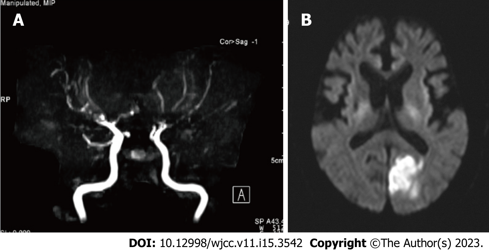
Figure 1 Magnetic resonance angiography and diffusion-weighted magnetic resonance imaging findings of Case 1.
A: Magnetic resonance angiography indicated the main branches of the internal carotid artery, including the middle cerebral artery, anterior cerebral artery, and vertebral artery, were tapered or disrupted; B: Diffusion-weighted magnetic resonance imaging indicates left occipital cerebral infarction.
- Citation: Harigane Y, Morimoto I, Suzuki O, Temmoku J, Sakamoto T, Nakamura K, Machii K, Miyata M. Enzyme replacement therapy in two patients with classic Fabry disease from the same family tree: Two case reports. World J Clin Cases 2023; 11(15): 3542-3551
- URL: https://www.wjgnet.com/2307-8960/full/v11/i15/3542.htm
- DOI: https://dx.doi.org/10.12998/wjcc.v11.i15.3542


