Copyright
©The Author(s) 2023.
World J Clin Cases. May 16, 2023; 11(14): 3340-3350
Published online May 16, 2023. doi: 10.12998/wjcc.v11.i14.3340
Published online May 16, 2023. doi: 10.12998/wjcc.v11.i14.3340
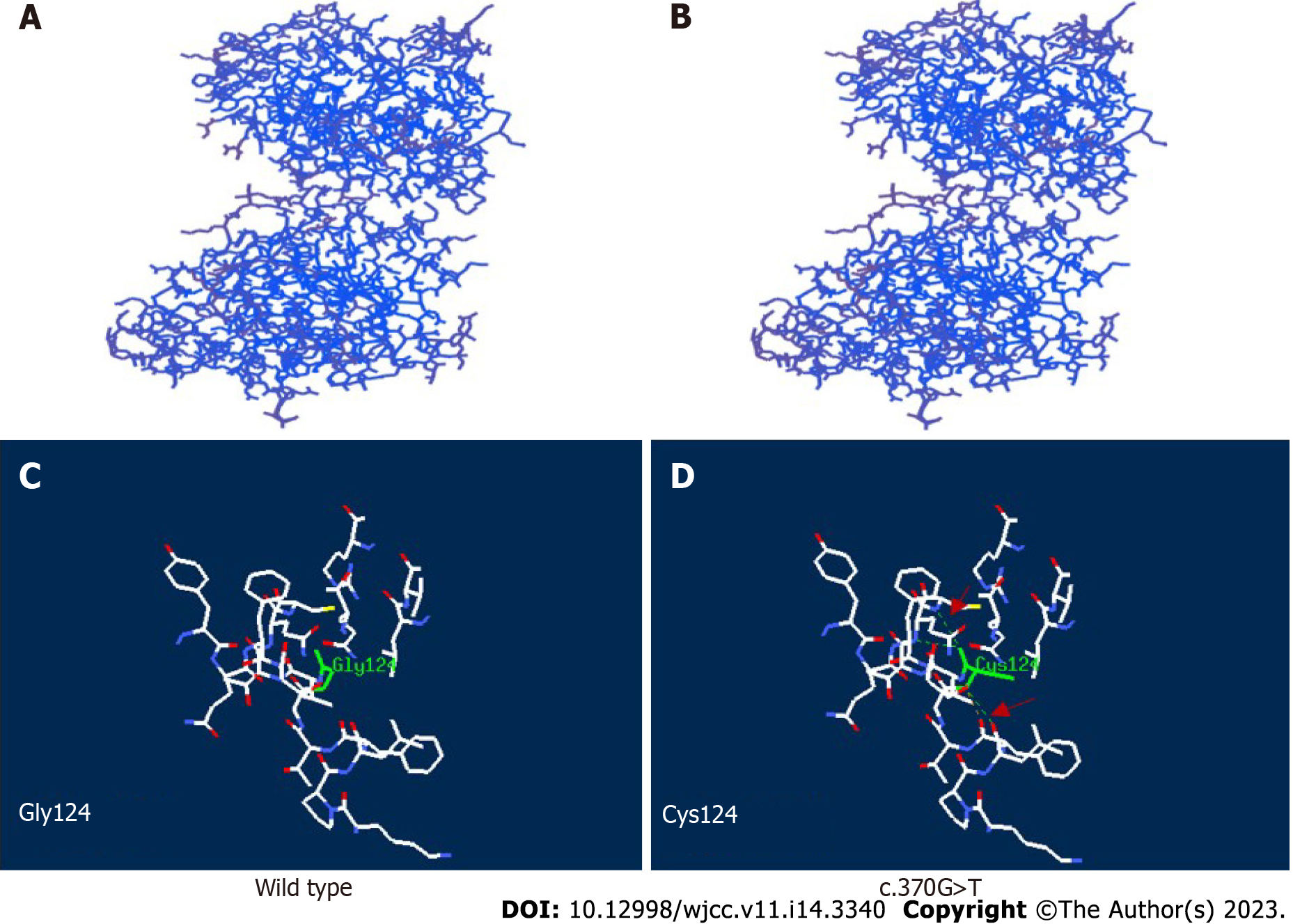
Figure 5 Structural analysis of wild-type and variant APOA1BP with c.
370G>T mutation. A: The three-dimensional structure of wild-type (WT); B: The three-dimensional structure of the mutant; C: The residue of missense mutant site together with the nearby functional site of WT; D: The residue of missense mutant site together with the nearby functional site of the mutant. Residues of the mutant sites are highlighted in green solid line. The computed hydrogen bonds are shown as green dashed lines and red arrow.
- Citation: Ding L, Huang TT, Ying GH, Wang SY, Xu HF, Qian H, Rahman F, Lu XP, Guo H, Zheng G, Zhang G. De novo mutation of NAXE (APOAIBP)-related early-onset progressive encephalopathy with brain edema and/or leukoencephalopathy-1: A case report. World J Clin Cases 2023; 11(14): 3340-3350
- URL: https://www.wjgnet.com/2307-8960/full/v11/i14/3340.htm
- DOI: https://dx.doi.org/10.12998/wjcc.v11.i14.3340


