Copyright
©The Author(s) 2023.
World J Clin Cases. May 16, 2023; 11(14): 3261-3266
Published online May 16, 2023. doi: 10.12998/wjcc.v11.i14.3261
Published online May 16, 2023. doi: 10.12998/wjcc.v11.i14.3261
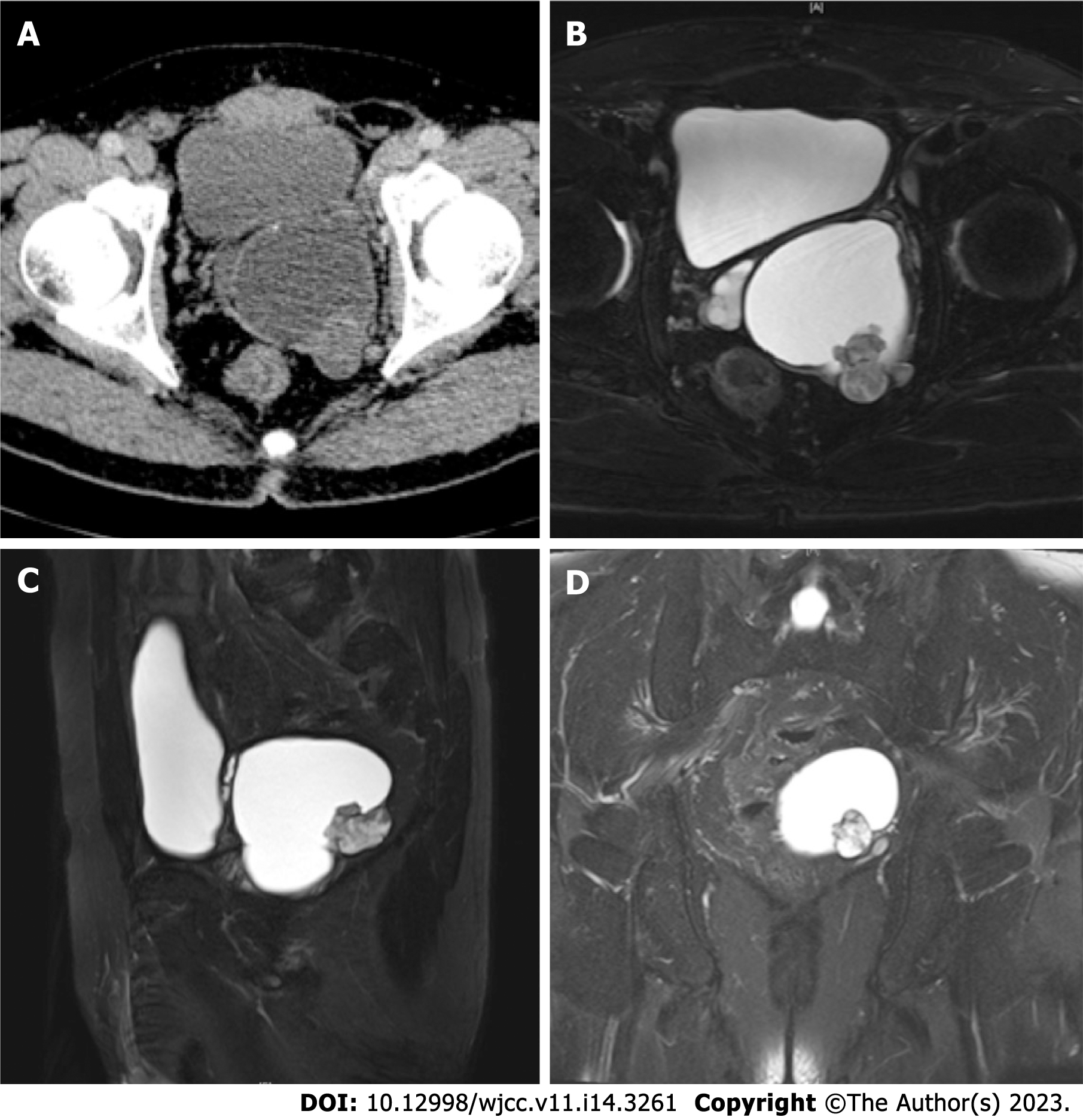
Figure 1 Imaging findings.
A: Axial of pelvic dynamic contrast-enhanced computed tomography revealed a cystic-solid mass. B-D: T2-weighted MRI imaging: Axial (B), sagittal (C), and coronal (D) sections of the pelvis demonstrated a cystic-solid component mass.
- Citation: Yao Y, Liu S, He YL, Luo L, Zhang GM. Primary seminal vesicle adenocarcinoma with a history of seminal vesicle cyst: A case report and review of literature. World J Clin Cases 2023; 11(14): 3261-3266
- URL: https://www.wjgnet.com/2307-8960/full/v11/i14/3261.htm
- DOI: https://dx.doi.org/10.12998/wjcc.v11.i14.3261


