Copyright
©The Author(s) 2023.
World J Clin Cases. May 6, 2023; 11(13): 3099-3104
Published online May 6, 2023. doi: 10.12998/wjcc.v11.i13.3099
Published online May 6, 2023. doi: 10.12998/wjcc.v11.i13.3099
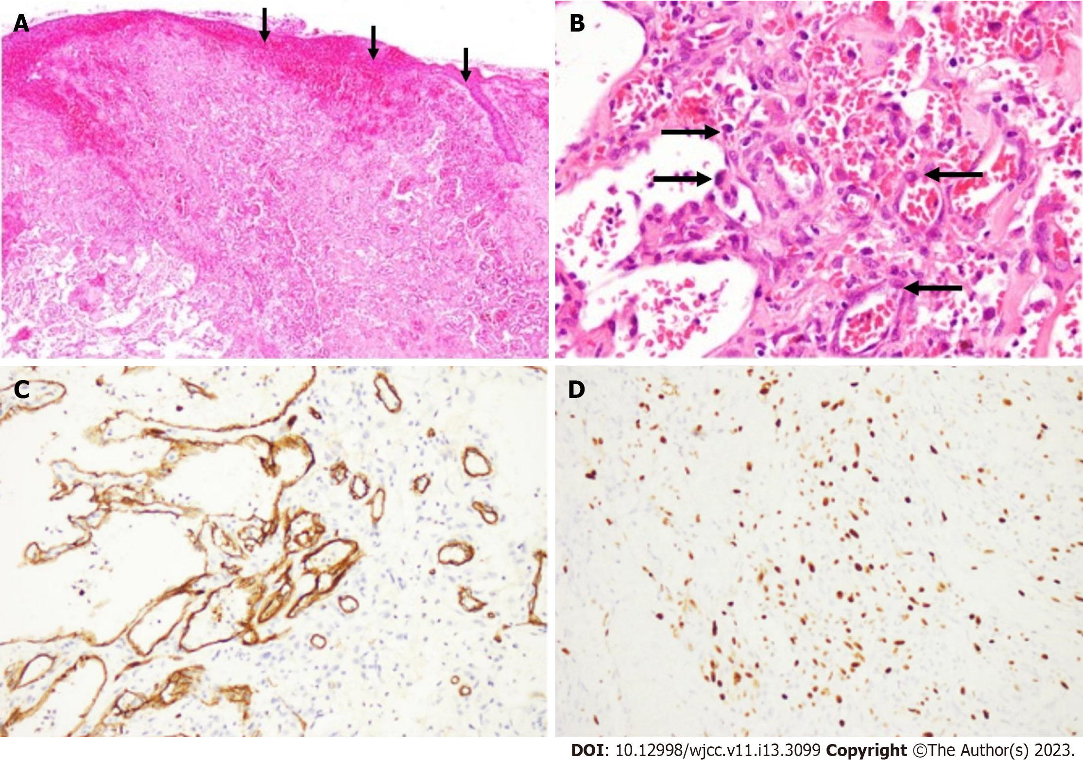
Figure 4 Morphological features of the angiosarcoma.
A: Under low magnification: The tumor cells diffusely infiltrated the dermis, involving the skin appendages and ulceration; B: Under high magnification: Proliferated vessels with fissures were composed of vascular endothelial cells with heteromorphic hyperplasia and nucleoli, and red blood cells were extravasated; C: Under high magnification: The intense positivity for an antiCD34 antibody shows that angiosarcomatous cells formed irregular vessels; D: Under high magnification: A high mitotic index was confirmed by immunohistochemical study for Ki67.
- Citation: Yan ZH, li ZL, Chen XW, Lian YW, Liu LX, Duan HY. Misdiagnosis of scalp angiosarcoma: A case report. World J Clin Cases 2023; 11(13): 3099-3104
- URL: https://www.wjgnet.com/2307-8960/full/v11/i13/3099.htm
- DOI: https://dx.doi.org/10.12998/wjcc.v11.i13.3099


