Copyright
©The Author(s) 2023.
World J Clin Cases. May 6, 2023; 11(13): 3076-3085
Published online May 6, 2023. doi: 10.12998/wjcc.v11.i13.3076
Published online May 6, 2023. doi: 10.12998/wjcc.v11.i13.3076
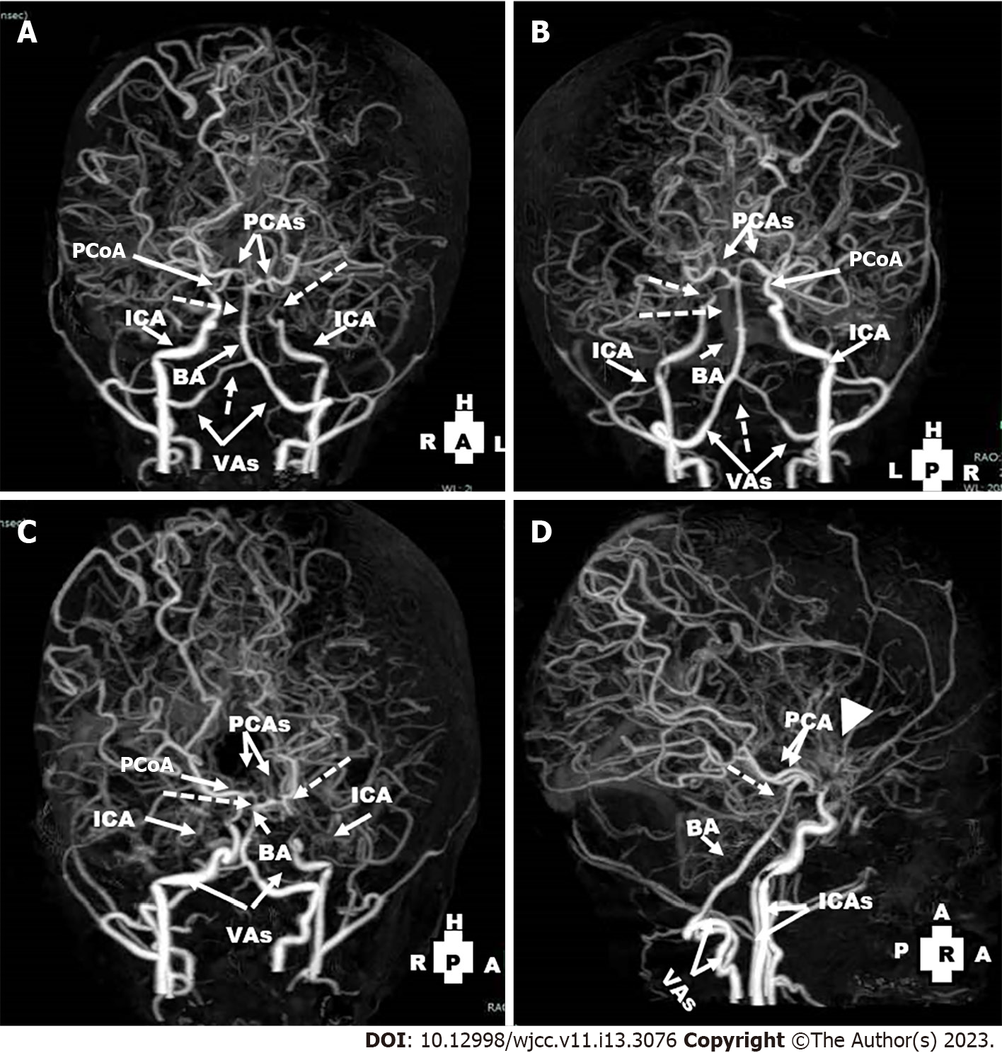
Figure 5 Computed tomography angiography views of the cerebral vessels (Date: January 2021): They revealed non-visualization of right and left anterior cerebral arteries and middle cerebral arteries.
A-C: The left internal carotid artery ended blindly before the siphon (dotted arrow), non-visualization of the left posterior communicating artery) and attenuation of the distal part of the right vertebral arteries and basilar artery (dotted arrows); D: There were extensive bilateral collaterals and prominence of moyamoya vessels (arrow head). PCoA: The posterior communicating artery; PCAs: The posterior cerebral arteries; ICA: The internal carotid artery; BA: The basilar artery; VAs: The vertebral arteries.
- Citation: Hamed SA, Yousef HA. Idiopathic steno-occlusive disease with bilateral internal carotid artery occlusion: A Case Report. World J Clin Cases 2023; 11(13): 3076-3085
- URL: https://www.wjgnet.com/2307-8960/full/v11/i13/3076.htm
- DOI: https://dx.doi.org/10.12998/wjcc.v11.i13.3076


