Copyright
©The Author(s) 2023.
World J Clin Cases. May 6, 2023; 11(13): 3076-3085
Published online May 6, 2023. doi: 10.12998/wjcc.v11.i13.3076
Published online May 6, 2023. doi: 10.12998/wjcc.v11.i13.3076
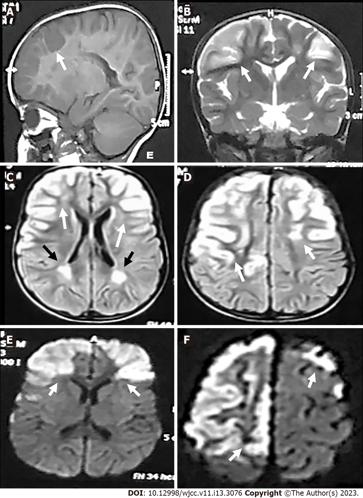
Figure 1 Magnetic resonance imaging-brain views (Date: July 2019).
A: Sagittal T1-weighted imaging revealed hypointense lesions in the fronto-parietal regions (white arrow); B: Coronal T2-weighted imaging; C and D: Axial fluid attenuation inversion recovery; E and F: Diffusion weighted imaging. Revealed hyperintense lesions in the right and left fronto-parietal areas (gyral pattern) (white arrows) and in the white matter adjacent to the lateral ventricle (C and D) (black arrows).
- Citation: Hamed SA, Yousef HA. Idiopathic steno-occlusive disease with bilateral internal carotid artery occlusion: A Case Report. World J Clin Cases 2023; 11(13): 3076-3085
- URL: https://www.wjgnet.com/2307-8960/full/v11/i13/3076.htm
- DOI: https://dx.doi.org/10.12998/wjcc.v11.i13.3076


