Copyright
©The Author(s) 2023.
World J Clin Cases. May 6, 2023; 11(13): 3038-3044
Published online May 6, 2023. doi: 10.12998/wjcc.v11.i13.3038
Published online May 6, 2023. doi: 10.12998/wjcc.v11.i13.3038
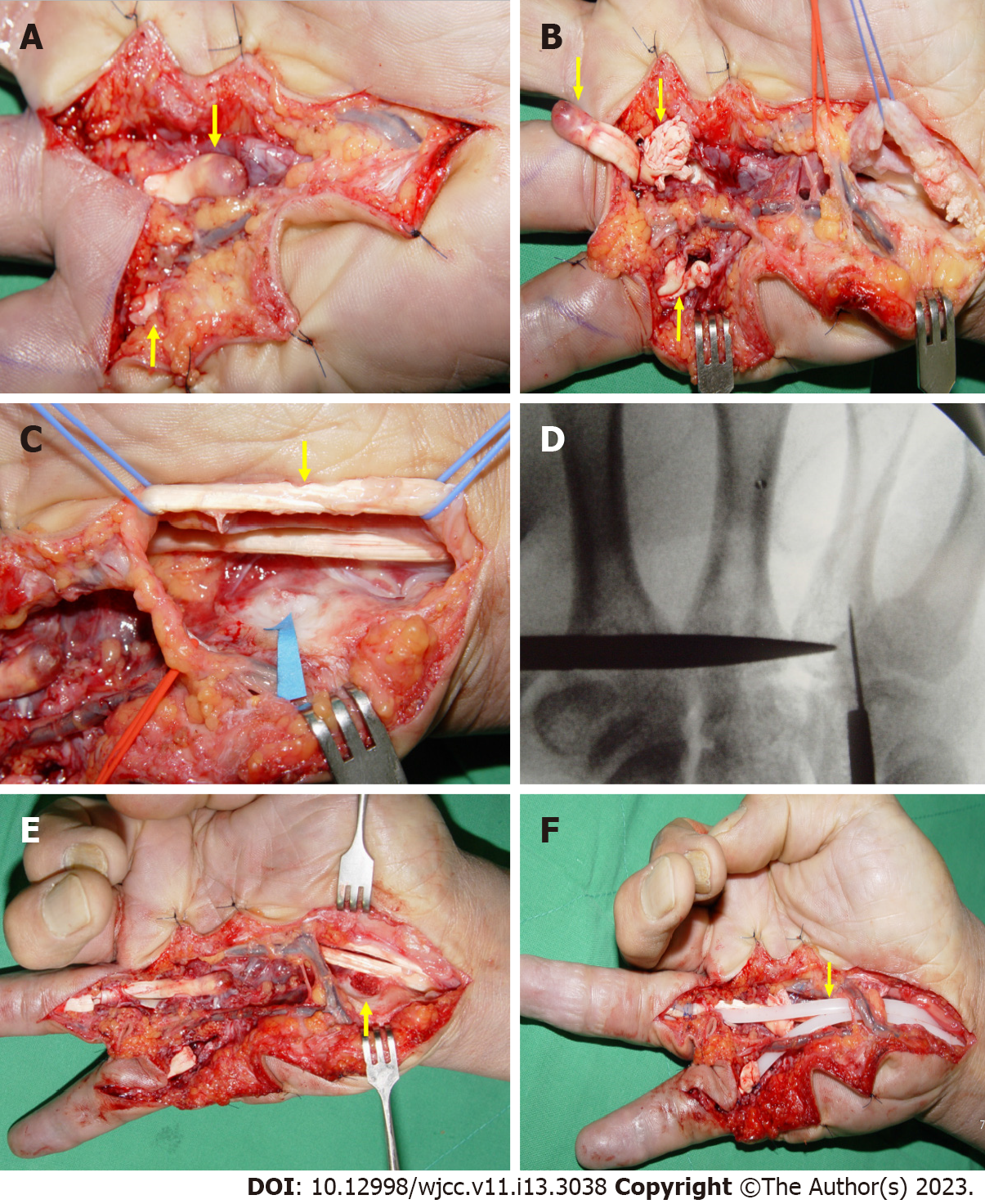
Figure 3 Intraoperative photographs.
A and B: Complete rupture of the right ring finger and right small finger flexor tendons (yellow arrows); C: A protruding bony structure like an osteophyte is identified on the side of the hamate in the carpal tunnel, and is covered by cartilage (blue arrow). There is a partial rupture of the right long finger flexor tendon (yellow arrow); D: The intra operative C-arm image shows a bony lesion protruding into the carpal tunnel; E: Excisional biopsy was performed for the bony lesion (yellow arrow); F: Hunter rods were inserted (yellow arrow).
- Citation: Kwon TY, Lee YK. Multiple flexor tendon ruptures due to osteochondroma of the hamate: A case report. World J Clin Cases 2023; 11(13): 3038-3044
- URL: https://www.wjgnet.com/2307-8960/full/v11/i13/3038.htm
- DOI: https://dx.doi.org/10.12998/wjcc.v11.i13.3038


