Copyright
©The Author(s) 2023.
World J Clin Cases. May 6, 2023; 11(13): 3010-3016
Published online May 6, 2023. doi: 10.12998/wjcc.v11.i13.3010
Published online May 6, 2023. doi: 10.12998/wjcc.v11.i13.3010
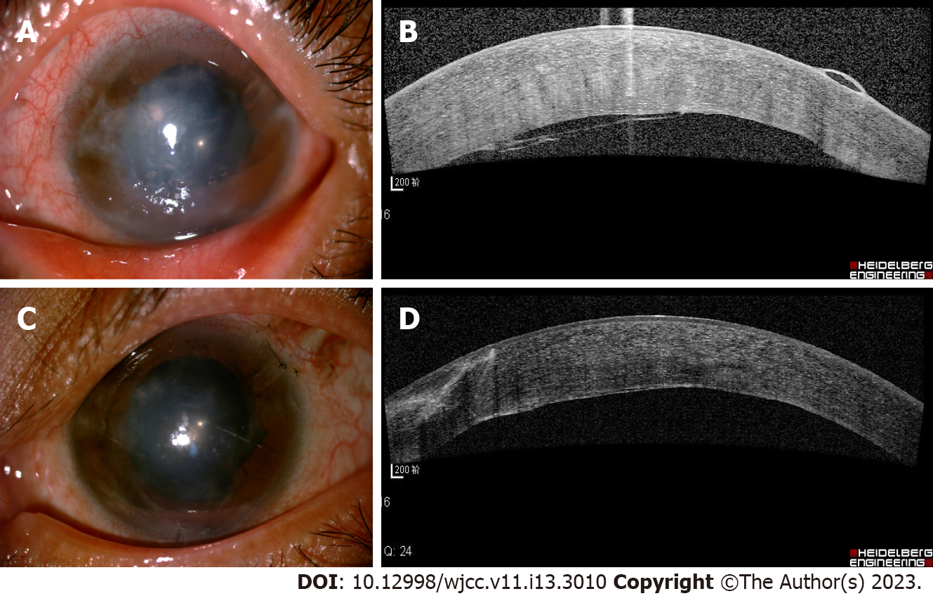
Figure 3 Anterior segment photography and optical coherence tomography of the left eye.
A: After peripheral iridectomy with zonulo-capsulo-hyaloidotomy and tension suture fixation, the anterior chamber was reconstructed. However, a significant corneal edema and bullous changes were seen; B: Optical coherence tomography showed that extensive corneal endothelial detachment involving approximately half of the cornea and a translucent membrane attached and stretched the endothelial layer; C: Two weeks after yttrium-aluminum-garnet laser, the corneal edema had abated, the corneal bullae had disappeared, and the peripheral cornea was transparent. D: Optical coherence tomography showed the corneal endothelial detachment gradually resolved. A highly reflective membranous mass attached to the medial endothelial layer.
- Citation: Ma YB, Dang YL. Bilateral malignant glaucoma with bullous keratopathy: A case report. World J Clin Cases 2023; 11(13): 3010-3016
- URL: https://www.wjgnet.com/2307-8960/full/v11/i13/3010.htm
- DOI: https://dx.doi.org/10.12998/wjcc.v11.i13.3010


