Copyright
©The Author(s) 2023.
World J Clin Cases. Apr 26, 2023; 11(12): 2839-2847
Published online Apr 26, 2023. doi: 10.12998/wjcc.v11.i12.2839
Published online Apr 26, 2023. doi: 10.12998/wjcc.v11.i12.2839
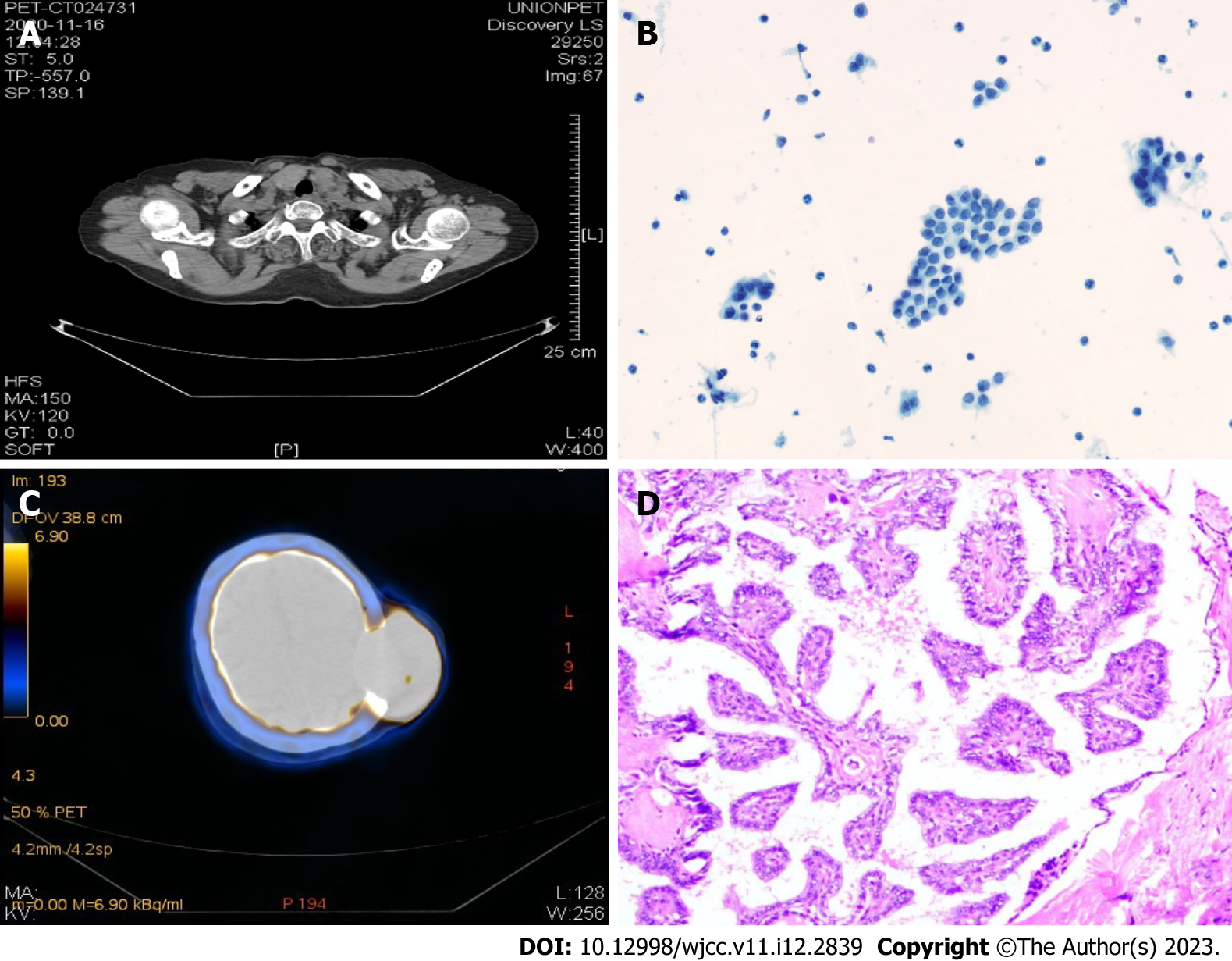
Figure 2 Histological and computed tomography images of the skull tumor.
A: positron emission tomography-computed tomography (PET-CT) imaging of the thyroid performed on initial diagnosis of the patient (November 16, 2020); B: Pathology of scalp mass biopsy (November 3, 2020). Scalp tumor histology similar to papillary thyroid carcinoma; papillary thyroid carcinoma metastasis was considered; C: PET-CT imaging of scalp metastasis (November 16, 2020). Note the hypermetabolic mass in the left temporoparietal bone, thinning of the corresponding temporoparietal bone due to compression, and local disappearance of the cerebral sulcus on the left side; D: Postoperative thyroid nodule pathology (paraffin) (March 18, 2021). Papillary thyroid carcinoma, classical type, interstitial fibrosis, no nerve infiltration or blood vessel invasion were observed. The tumor involved extra-glandular fibrofatty tissue.
- Citation: Zhang LY, Cai SJ, Liang BY, Yan SY, Wang B, Li MY, Zhao WX. Efficacy of anlotinib combined with radioiodine to treat scalp metastasis of papillary thyroid cancer: A case report and review of literature. World J Clin Cases 2023; 11(12): 2839-2847
- URL: https://www.wjgnet.com/2307-8960/full/v11/i12/2839.htm
- DOI: https://dx.doi.org/10.12998/wjcc.v11.i12.2839


